Dept. of Radiology, Feinberg School of Medicine, Northwestern University, 737 N. Michigan Ave, Suite 1600, Chicago, IL, 60611, USA.
Dept. of Gastrointestinal Surgery, Affiliated Hospital of Qingdao University, Qingdao, Shandong, China.
J Transl Med. 2020 Feb 10;18(1):61. doi: 10.1186/s12967-020-02246-7.
There is a lack of well-established clinical tools for predicting dendritic cell (DC) vaccination response of pancreatic ductal adenocarcinoma (PDAC). DC vaccine treatment efficiency was demonstrated using histological analysis in pre-clinical studies; however, its usage was limited due to invasiveness. In this study, we aimed to investigate the potential of MRI texture features for detection of early immunotherapeutic response as well as overall survival (OS) of PDAC subjects following dendritic cell (DC) vaccine treatment in LSL-Kras;LSL-Trp53;Pdx-1-Cre (KPC) transgenic mouse model of pancreatic ductal adenocarcinoma (PDAC).
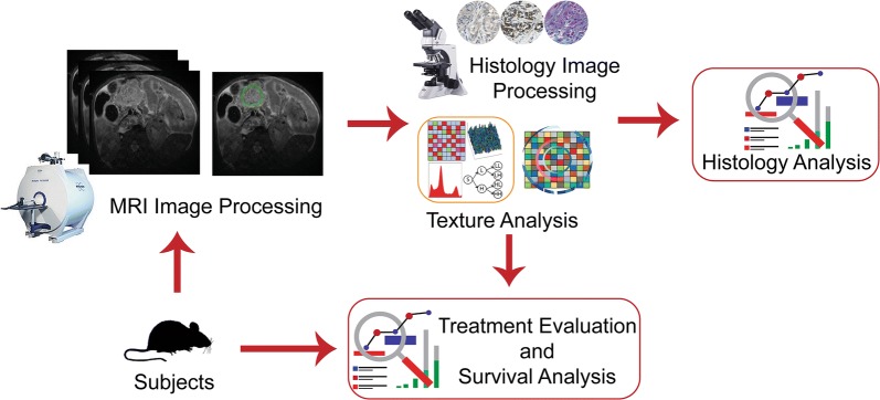
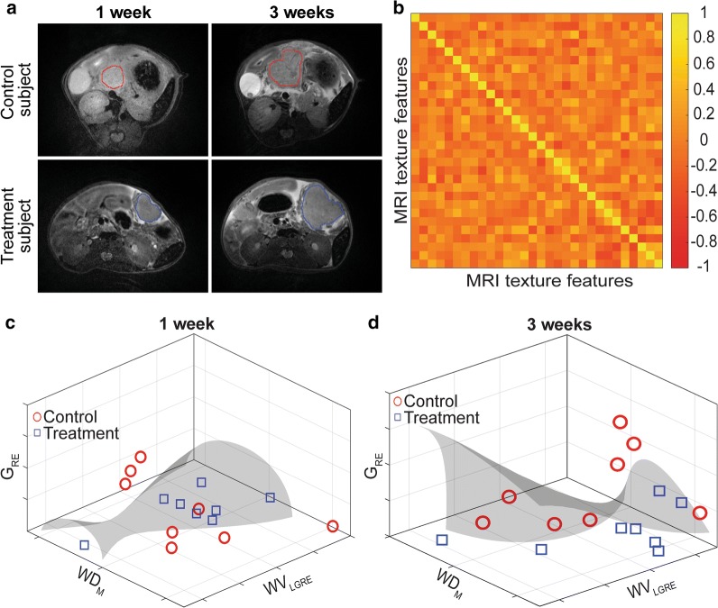
KPC mice were treated with DC vaccines, and tumor growth was dynamically monitored. A total of a hundred and fifty-two image features of T2-weighted MRI images were analyzed using a kernel-based support vector machine model to detect treatment effects following the first and third weeks of the treatment. Moreover, univariate analysis was performed to describe the association between MRI texture and survival of KPC mice as well as histological tumor biomarkers.
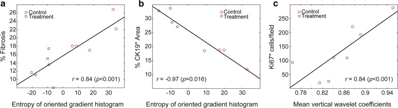
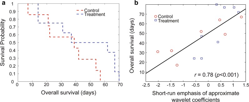
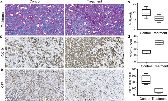
OS for mice in the treatment group was 54.8 ± 22.54 days while the control group had 35.39 ± 17.17 days. A subset of three MRI features distinguished treatment effects starting from the first week with increasing accuracy throughout the treatment (75% to 94%). Besides, we observed that short-run emphasis of approximate wavelet coefficients had a positive correlation with the survival of the KPC mice (r = 0.78, p < 0.001). Additionally, tissue-specific MRI texture features showed positive association with fibrosis percentage (r = 0.84, p < 0.002), CK19 positive percentage (r = - 0.97, p < 0.001), and Ki67 positive cells (r = 0.81, p < 0.02) as histological disease biomarkers.
Our results demonstrate that MRI texture features can be used as imaging biomarkers for early detection of therapeutic response following DC vaccination in the KPC mouse model of PDAC. Besides, MRI texture can be utilized to characterize tumor microenvironment reflected with histology analysis.
目前缺乏用于预测胰腺导管腺癌(PDAC)树突状细胞(DC)疫苗反应的成熟临床工具。在临床前研究中,通过组织学分析证明了 DC 疫苗治疗的效率;然而,由于其侵入性,其使用受到限制。在这项研究中,我们旨在研究 MRI 纹理特征在检测 LSL-Kras;LSL-Trp53;Pdx-1-Cre(KPC)转基因小鼠胰腺导管腺癌(PDAC)模型中接受树突状细胞(DC)疫苗治疗后 PDAC 患者早期免疫治疗反应和总生存期(OS)的潜力。
KPC 小鼠接受 DC 疫苗治疗,并动态监测肿瘤生长。使用基于核的支持向量机模型分析 T2 加权 MRI 图像的 152 个图像特征,以检测治疗后第一周和第三周的治疗效果。此外,进行单变量分析以描述 MRI 纹理与 KPC 小鼠生存以及组织学肿瘤标志物之间的关系。
治疗组小鼠的 OS 为 54.8±22.54 天,而对照组为 35.39±17.17 天。从第一周开始,有一组三个 MRI 特征可以区分治疗效果,并且随着治疗的进行,准确性逐渐提高(75%至 94%)。此外,我们观察到短程近似小波系数的重点与 KPC 小鼠的生存呈正相关(r=0.78,p<0.001)。此外,组织特异性 MRI 纹理特征与纤维化百分比(r=0.84,p<0.002)、CK19 阳性百分比(r=-0.97,p<0.001)和 Ki67 阳性细胞(r=0.81,p<0.02)呈正相关作为组织学疾病标志物。
我们的结果表明,MRI 纹理特征可用作 PDAC KPC 小鼠模型中 DC 疫苗治疗后早期检测治疗反应的成像生物标志物。此外,MRI 纹理可用于描绘与组织学分析相关的肿瘤微环境。