Shafqat Muhammad Nabeel, Memon Muhammad Yousuf Y, Javed Salman, Kanagala Sai Gautham, Saleem Momina
Department of Gastroenterology and Hepatology, Allied Teaching Hospital Gujranwala, Gujranwala, PAK.
Department of Gastroenterology, King Saud Hospital, Unaizah, SAU.
Cureus. 2023 Dec 20;15(12):e50871. doi: 10.7759/cureus.50871. eCollection 2023 Dec.
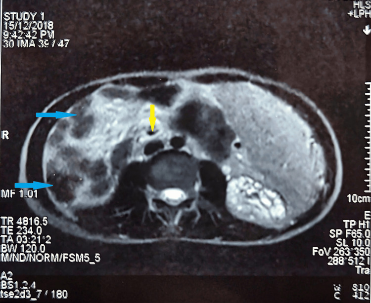
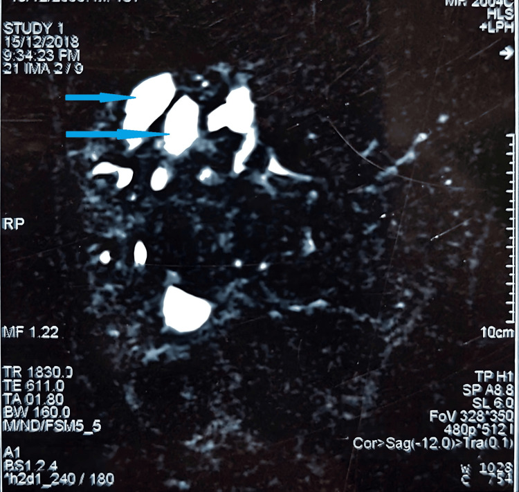
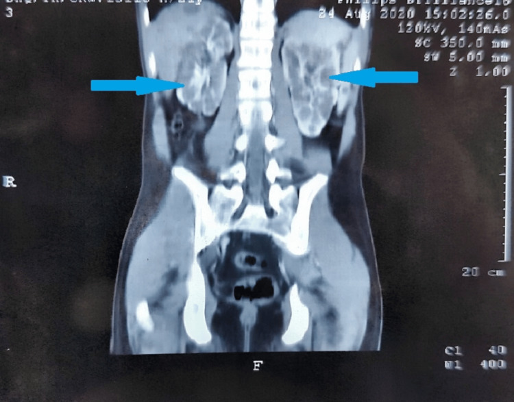
Synonymous with congenital non-obstructive saccular or fusiform intra-hepatic duct dilatation and congenital communicating cavernous ectasia of the intra-hepatic biliary tract, Caroli's syndrome (CS) is an extremely rare fibro-polycystic liver disorder characterized by ductal plate malformation and consequent peri-portal fibrosis due to segmental intra-hepatic duct dilatation. No more than 200 cases of the syndrome have been reported since 1958. CS may affect one or both lobes of the liver, but more commonly it affects the left hepatic lobe. We describe a rare case of CS localized to the right hepatic lobe in a 21-year-old male, who presented with complaints of upper gastrointestinal (GI) bleeding without any signs or stigmata of chronic liver disease. Personal as well as family history was non-significant except positive for consanguineous parental marriage. General physical examination was unremarkable except for pallor, and upper GI endoscopy revealed columns of bandable esophageal varices which led us to a line of investigations to identify the cause of portal hypertension. Blood tests were non-specific, though imaging studies chiefly abdominal ultrasound, CT abdomen and pelvis with contrast, and magnetic resonance cholangiopancreatography (MRCP) led us to confirmation of the diagnosis of CS in the right hepatic lobe with manifestations of portal hypertension as the predominant feature. Diagnosis was confirmed on liver biopsy which showed right-sided cystic dilations with congenital hepatic fibrosis.
卡罗里氏综合征(CS)与先天性非梗阻性囊状或梭形肝内胆管扩张以及先天性肝内胆管交通性海绵状扩张同义,是一种极为罕见的纤维多囊性肝脏疾病,其特征为导管板畸形以及由于节段性肝内胆管扩张导致的门静脉周围纤维化。自1958年以来,该综合征的报告病例不超过200例。CS可累及肝脏的一叶或两叶,但更常见的是累及左肝叶。我们描述了一例罕见的CS病例,病变局限于一名21岁男性的右肝叶,该患者表现为上消化道(GI)出血,且无任何慢性肝病的体征或迹象。除近亲父母婚姻史呈阳性外,个人及家族史均无异常。除面色苍白外,全身体格检查无异常,上消化道内镜检查发现可套扎的食管静脉曲张柱,这使我们展开一系列检查以确定门静脉高压的病因。血液检查无特异性,尽管影像学检查主要包括腹部超声、腹部和盆腔增强CT以及磁共振胰胆管造影(MRCP),这些检查使我们确诊右肝叶CS,并以门静脉高压表现为主要特征。肝活检证实了诊断,显示右侧囊性扩张并伴有先天性肝纤维化。