Department of Biomedical Systems Informatics (J.H., Y.R.P.), Yonsei University College of Medicine, Seoul, Republic of Korea.
Department of Medical Humanities and Social Sciences (S.Y.), Yonsei University College of Medicine, Seoul, Republic of Korea.
Stroke. 2024 Mar;55(3):715-724. doi: 10.1161/STROKEAHA.123.044026. Epub 2024 Jan 23.
Moyamoya disease (MMD) is a rare and complex pathological condition characterized by an abnormal collateral circulation network in the basal brain. The diagnosis of MMD and its progression is unpredictable and influenced by many factors. MMD can affect the blood vessels supplying the eyes, resulting in a range of ocular symptoms. In this study, we developed a deep learning model using real-world data to assist a diagnosis and determine the stage of the disease using retinal photographs.
This retrospective observational study conducted from August 2006 to March 2022 included 498 retinal photographs from 78 patients with MMD and 3835 photographs from 1649 healthy participants. Photographs were preprocessed, and an ResNeXt50 model was developed. Model performance was measured using receiver operating curves and their area under the receiver operating characteristic curve, accuracy, sensitivity, and F1-score. Heatmaps and progressive erasing plus progressive restoration were performed to validate the faithfulness.
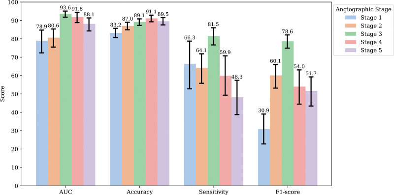
Overall, 322 retinal photographs from 67 patients with MMD and 3752 retinal photographs from 1616 healthy participants were used to develop a screening and stage prediction model for MMD. The average age of the patients with MMD was 44.1 years, and the average follow-up time was 115 months. Stage 3 photographs were the most prevalent, followed by stages 4, 5, 2, 1, and 6 and healthy. The MMD screening model had an average area under the receiver operating characteristic curve of 94.6%, with 89.8% sensitivity and 90.4% specificity at the best cutoff point. MMD stage prediction models had an area under the receiver operating characteristic curve of 78% or higher, with stage 3 performing the best at 93.6%. Heatmap identified the vascular region of the fundus as important for prediction, and progressive erasing plus progressive restoration result shows an area under the receiver operating characteristic curve of 70% only with 50% of the important regions.
This study demonstrated that retinal photographs could be used as potential biomarkers for screening and staging of MMD and the disease stage could be classified by a deep learning algorithm.
烟雾病(MMD)是一种罕见且复杂的病理状况,其特征在于基底脑内的异常侧支循环网络。MMD 的诊断及其进展是不可预测的,受到许多因素的影响。MMD 可影响供应眼睛的血管,导致一系列眼部症状。在这项研究中,我们使用真实世界的数据开发了一个深度学习模型,以协助诊断并使用视网膜照片确定疾病的阶段。
这项回顾性观察研究于 2006 年 8 月至 2022 年 3 月进行,纳入了 78 例 MMD 患者的 498 张视网膜照片和 1649 例健康参与者的 3835 张照片。对照片进行预处理,并开发了 ResNeXt50 模型。使用接收者操作曲线及其曲线下面积、准确性、敏感性和 F1 分数来衡量模型性能。进行热图和逐步擦除加逐步恢复以验证忠实度。
总体而言,我们使用 67 例 MMD 患者的 322 张视网膜照片和 1616 例健康参与者的 3752 张视网膜照片开发了用于 MMD 筛查和分期预测的模型。MMD 患者的平均年龄为 44.1 岁,平均随访时间为 115 个月。3 期照片最常见,其次是 4 期、5 期、2 期、1 期和 6 期以及健康者。MMD 筛查模型的平均接收者操作特征曲线下面积为 94.6%,最佳截止点的敏感性为 89.8%,特异性为 90.4%。MMD 分期预测模型的接收者操作特征曲线下面积在 78%或更高,其中 3 期的表现最佳,为 93.6%。热图确定眼底的血管区域对预测很重要,逐步擦除加逐步恢复结果显示,仅在 50%的重要区域存在时,接收者操作特征曲线下面积为 70%。
本研究表明,视网膜照片可用作 MMD 的筛查和分期的潜在生物标志物,并且可以通过深度学习算法对疾病阶段进行分类。