College of Veterinary Medicine, Hunan Agricultural University, 410128, Changsha, Hunan, China.
College of Veterinary Medicine, Northwest A&F University, 712100, Yangling, Shaanxi, China.
Virol J. 2024 Jan 23;21(1):25. doi: 10.1186/s12985-024-02289-y.
Pseudorabies virus (PRV) is one of the major viral pathogens leading to reproductive disorders in swine. However, little is known about the effects of PRV infection on porcine reproductive system. Ovarian granulosa cells are somatic cells surrounding oocytes in ovary and required for folliculogenesis. The present study aimed to investigate the interference of PRV on functions of porcine ovarian granulosa cells in vitro.
Primary granulosa cells were isolated from porcine ovaries. To investigate the PRV infectivity, transmission electron microscopy (TEM) was used to check the presence of viral particles, and the expression of viral gE gene was detected by quantitative real-time PCR (qPCR) in PRV-inoculated cells. After PRV infection, cell viability was detected by MTS assay, Ki67 for proliferative status was determined by immunofluorescence assay (IFA), cell cycle and apoptosis were detected by flow cytometry, and progesterone (P) and estradiol (E) were determined by radioimmunoassay. The checkpoint genes of cell cycle and apoptosis-related proteins were studied by qPCR and western blotting.
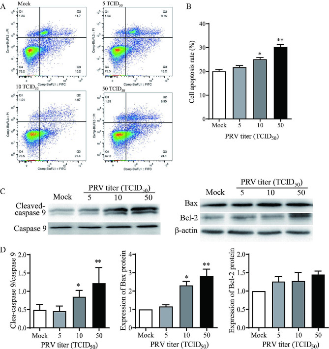
Virus particles were observed in the nucleus and cytoplasm of PRV-infected granulosa cells by TEM imaging, and the expression of viral gE gene increased in a time-dependent manner post infection. PRV infection inhibited cell viability and blocked cell cycle at S phase in porcine granulosa cells, accompanied by decreases in expression of Ki67 protein and checkpoint genes related to S phase. Radioimmunoassay revealed decreased levels in P and E, and the expressions of key steroidogenic enzymes were also down-regulated post PRV-infection. In addition, PRV induced apoptosis with an increase in Bax expression and activation of caspase 9, and the phosphorylation of JNK, ERK and p38 MAPKs were significantly up-regulated in porcine ovarian granulosa cells post PRV infection.
The data indicate that PRV causes infection on porcine ovarian granulosa cells and interferes the cell functions through apoptosis, and the MAPK signaling pathway is involved in the viral pathogenesis.
伪狂犬病毒(PRV)是导致猪繁殖障碍的主要病毒病原体之一。然而,关于 PRV 感染对猪生殖系统的影响知之甚少。卵巢颗粒细胞是环绕卵巢中卵母细胞的体细胞,是卵泡发生所必需的。本研究旨在探讨 PRV 在体外对猪卵巢颗粒细胞功能的干扰。
从猪卵巢中分离原代颗粒细胞。为了研究 PRV 的感染性,使用透射电子显微镜(TEM)检查病毒颗粒的存在,并通过定量实时 PCR(qPCR)检测 PRV 感染细胞中病毒 gE 基因的表达。PRV 感染后,通过 MTS 测定法检测细胞活力,通过免疫荧光法(IFA)测定 Ki67 增殖状态,通过流式细胞术检测细胞周期和凋亡,通过放射免疫法测定孕酮(P)和雌二醇(E)。通过 qPCR 和 Western blot 研究细胞周期和凋亡相关蛋白的检查点基因。
TEM 成像观察到 PRV 感染的颗粒细胞的核和细胞质中存在病毒颗粒,感染后病毒 gE 基因的表达呈时间依赖性增加。PRV 感染抑制猪颗粒细胞的活力并在 S 期阻断细胞周期,伴随 Ki67 蛋白和与 S 期相关的检查点基因表达减少。放射免疫法显示 P 和 E 水平降低,PRV 感染后关键类固醇生成酶的表达也下调。此外,PRV 诱导细胞凋亡,Bax 表达增加,caspase 9 激活,PRV 感染后猪卵巢颗粒细胞中 JNK、ERK 和 p38 MAPK 的磷酸化明显上调。
数据表明 PRV 引起猪卵巢颗粒细胞感染,并通过凋亡干扰细胞功能,MAPK 信号通路参与病毒发病机制。