Doheny Eye Institute, Pasadena, California, United States.
Department of Ophthalmology David Geffen School of Medicine, University of California Los Angeles, Los Angeles, California, United States.
Invest Ophthalmol Vis Sci. 2024 Jan 2;65(1):47. doi: 10.1167/iovs.65.1.47.
To compare optical coherence tomography angiography (OCTA) retina metrics between cognitively healthy subjects with pathological versus normal cerebrospinal fluid (CSF) Aβ42/tau ratios.
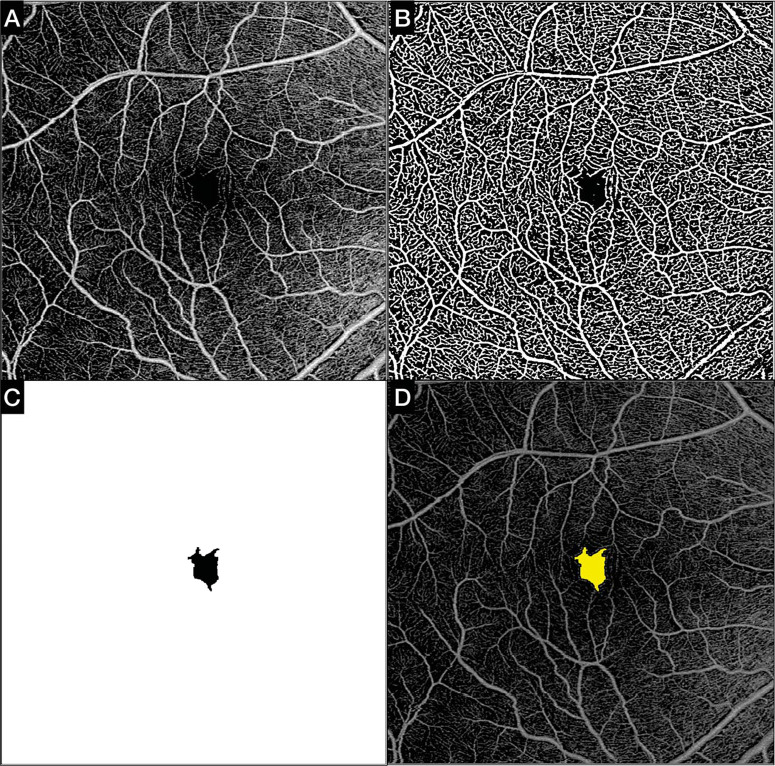
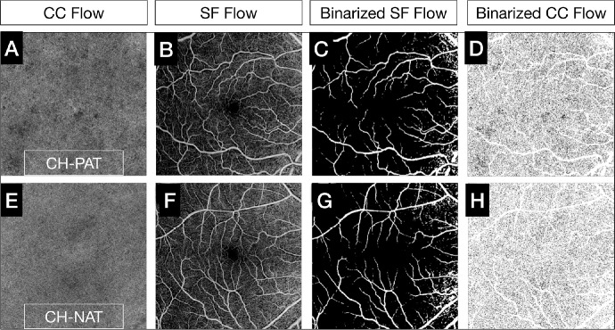
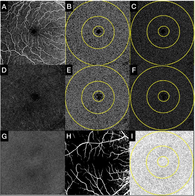
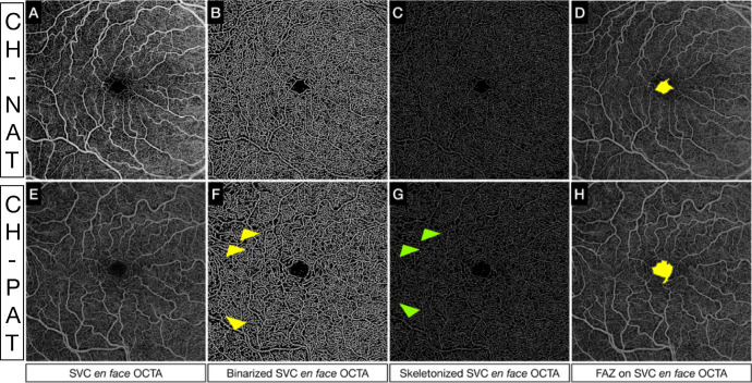
Swept-source OCTA scans were collected using the Zeiss PLEX Elite 9000 and analyzed on 23 cognitively healthy (CH) subjects who had previously undergone CSF analysis. Thirteen subjects had a pathological Aβ42/tau (PAT) ratio of <2.7132, indicative of presymptomatic Alzheimer's disease (AD), and 10 had a normal Aβ42/tau (NAT) ratio of ≥2.7132. OCTA en face images of the superficial vascular complex (SVC) and deep vascular complex were binarized and skeletonized to quantify the perfusion density (PD), vessel length density (VLD), and fractal dimension (FrD). The foveal avascular zone (FAZ) area was calculated using the SVC slab. Choriocapillaris flow deficits (CCFDs) were computed from the en face OCTA slab of the CC. The above parameters were compared between CH-PATs and CH-NATs.
Compared to CH-NATs, CH-PATs showed significantly decreased PD, VLD, and FrD in the SVC, with a significantly increased FAZ area and CCFDs.
Swept-source OCTA analysis of the SVC and CC suggests a significant vascular loss at the CH stage of pre-AD that might be an indicator of a neurodegenerative process initiated by the impaired clearance of Aβ42 in the blood vessel wall and by phosphorylated tau accumulation in the perivascular spaces, a process that most likely mirrors that in the brain. If confirmed in larger longitudinal studies, OCTA retinal and inner choroidal metrics may be important biomarkers for assessing presymptomatic AD.
比较认知健康受试者中脑脊液(CSF) Aβ42/τ比值病理性与正常的光学相干断层扫描血管造影(OCTA)视网膜指标。
使用 Zeiss PLEX Elite 9000 采集扫频源 OCTA 扫描,并对先前进行过 CSF 分析的 23 名认知健康(CH)受试者进行分析。13 名受试者的 Aβ42/τ(PAT)比值<2.7132,提示有前驱期阿尔茨海默病(AD),10 名受试者的 Aβ42/τ(NAT)比值≥2.7132。对浅层血管丛(SVC)和深层血管丛的 OCTA 共焦图像进行二值化和骨架化,以量化灌注密度(PD)、血管长度密度(VLD)和分形维数(FrD)。使用 SVC 层计算中央无血管区(FAZ)面积。从脉络膜毛细血管 OCTA 共焦层计算脉络膜毛细血管血流缺损(CCFDs)。比较 CH-PATs 和 CH-NATs 之间的上述参数。
与 CH-NATs 相比,CH-PATs 的 SVC 中 PD、VLD 和 FrD 显著降低,FAZ 面积显著增加,CCFDs 显著增加。
SVC 和 CC 的扫频源 OCTA 分析表明,在 AD 前驱期的 CH 阶段存在明显的血管丢失,这可能是血管壁 Aβ42 清除受损和血管周围空间磷酸化 tau 积聚引发的神经退行性过程的一个指标,这个过程很可能反映了大脑中的情况。如果在更大的纵向研究中得到证实,OCTA 视网膜和内层脉络膜指标可能是评估前驱期 AD 的重要生物标志物。