Hong Si-Jie, Hong Li-Wei, He Xiao-Qin, Zhong Xiao-Hong
Department of Ultrasound, Women and Children's Hospital, School of Medicine, Xiamen University, Xiamen 361000, Fujian Province, China.
Ministry of Science and Education, Women and Children's Hospital, School of Medicine, Xiamen University, Xiamen 361000, Fujian Province, China.
World J Clin Cases. 2024 Jan 16;12(2):240-248. doi: 10.12998/wjcc.v12.i2.240.
Umbilical artery thrombosis (UAT) is extremely uncommon and leads to adverse perinatal outcomes. Hypercoagulation of blood in pregnant women is suspected to be an important risk for UAT. Ultrasound is an effective way to detect thrombosis. The mother can monitor her own fetal health using ultrasound, which enables her to take preventative action in case of emergency.
To investigate ultrasonic blood signal after UAT in the umbilical artery, and evaluate the relationship between hypercoagulability and UAT.
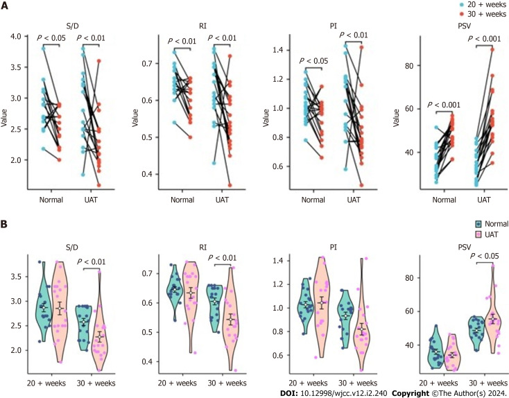
We described a case of a newly formed UAT with markedly altered ultrasonic indices of umbilical artery blood flow, and retrospectively studied it with 18 UAT patients confirmed by histopathology from October 2019 and March 2023 in Xiamen Women and Children's Hospital. Patients' information was collected from medical archives, including maternal clinical data, neonatal outcomes, pathological findings and ultrasonic indices of umbilical artery blood flow, such as systolic-diastolic duration ratio (S/D), resistance index (RI), pulsatility index (PI) and peak systolic velocity (PSV). Ultrasound and coagulation indices were analyzed with matched samples -test and Wilcoxon rank sum test using the statistical packages in R (version 4.2.1) including car (version 3.1-0) and stats (version 4.2.1), and visualized by ggplot2 package (version 3.3.6).
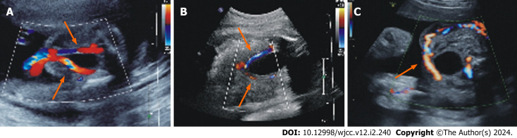
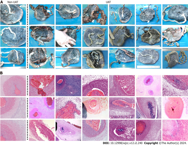
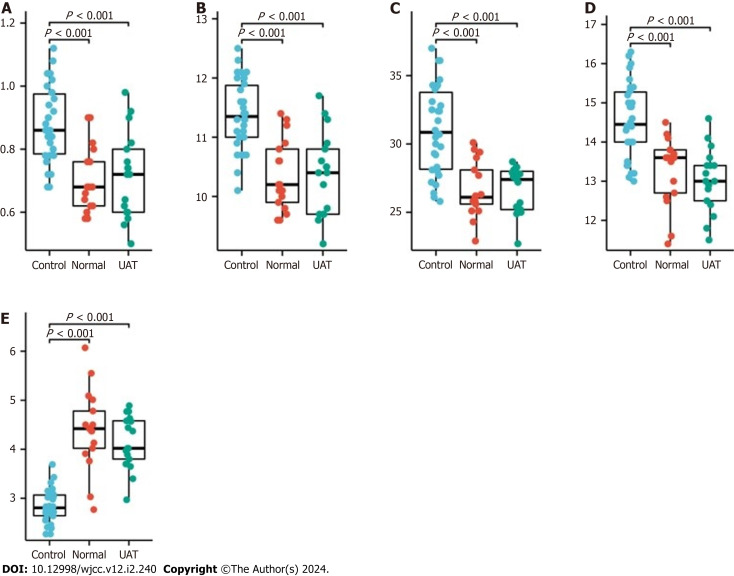
A patient with normal findings in second and third-trimester routine ultrasound scan developed UAT with severe changes in ultrasonic indices of umbilical artery blood flow (within 2.5 of reference ranges) in a short period of time. Statistical analysis of umbilical artery blood flow ultrasound indices for 19 patients with UAT showed that the decrease in S/D, RI, and PI and increase of PSV during the disease process was greater than that of non-UAT. All 18 patients delivered in our hospital showed characteristic manifestations of UAT on histological examination after delivery, most of which (16/18) showed umbilical cord abnormalities, with 15 umbilical cord torsion and 1 pseudoknot. Coagulation parameters were not significantly changed in UAT patients compared with normal pregnancy women.
Significant changes in ultrasound indicators after UAT were demonstrated. PSV can play important roles in the diagnosis of UAT. Hypercoagulability alone is not sufficient for the occurrence of UAT.
脐动脉血栓形成(UAT)极为罕见,并会导致不良围产期结局。孕妇血液高凝状态被怀疑是UAT的一个重要风险因素。超声是检测血栓形成的有效方法。母亲可以使用超声监测自身胎儿的健康状况,这使她能够在紧急情况下采取预防措施。
研究脐动脉发生UAT后的超声血流信号,并评估血液高凝状态与UAT之间的关系。
我们描述了1例新形成的UAT病例,其脐动脉血流超声指标有明显改变,并对2019年10月至2023年3月在厦门市妇幼保健院经组织病理学确诊的18例UAT患者进行回顾性研究。从医疗档案中收集患者信息,包括母亲的临床资料、新生儿结局、病理检查结果以及脐动脉血流超声指标,如收缩期与舒张期持续时间比(S/D)、阻力指数(RI)、搏动指数(PI)和收缩期峰值流速(PSV)。使用R(版本4.2.1)中的统计软件包,包括car(版本3.1 - 0)和stats(版本4.2.1),通过配对样本t检验和Wilcoxon秩和检验分析超声和凝血指标,并使用ggplot2软件包(版本3.3.6)进行可视化。
1例在孕中期和孕晚期常规超声检查结果正常的患者发生了UAT,脐动脉血流超声指标在短时间内出现严重变化(在参考范围的2.5倍以内)。对19例UAT患者的脐动脉血流超声指标进行统计分析显示,疾病过程中S/D、RI和PI的降低以及PSV的升高幅度大于非UAT患者。我院分娩的18例患者产后组织学检查均显示UAT的特征性表现,其中大部分(16 / 18)存在脐带异常,15例脐带扭转,1例假结。与正常孕妇相比,UAT患者的凝血参数无明显变化。
证实了UAT后超声指标有显著变化。PSV在UAT诊断中可发挥重要作用。单纯血液高凝状态不足以导致UAT的发生。