Wong Yin Ping, Abd Rahman Rahana, Tan Ay Eeng, Tan Geok Chin
Department of Pathology, Faculty of Medicine, Universiti Kebangsaan Malaysia, Kuala Lumpur 56000, Malaysia.
Department of Obstetrics and Gynaecology, Faculty of Medicine, Universiti Kebangsaan Malaysia, Kuala Lumpur 56000, Malaysia.
Diagnostics (Basel). 2025 Jan 3;15(1):94. doi: 10.3390/diagnostics15010094.
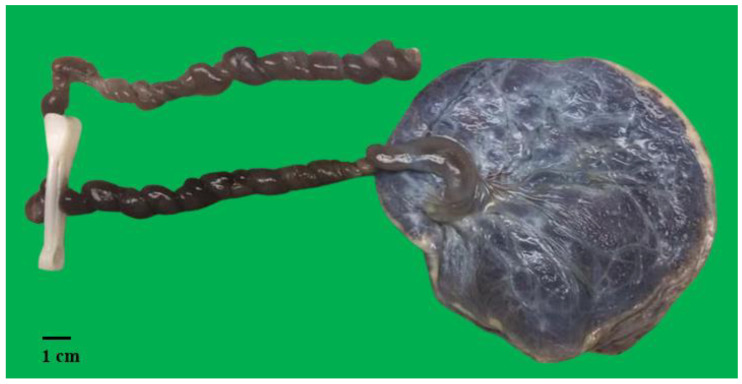
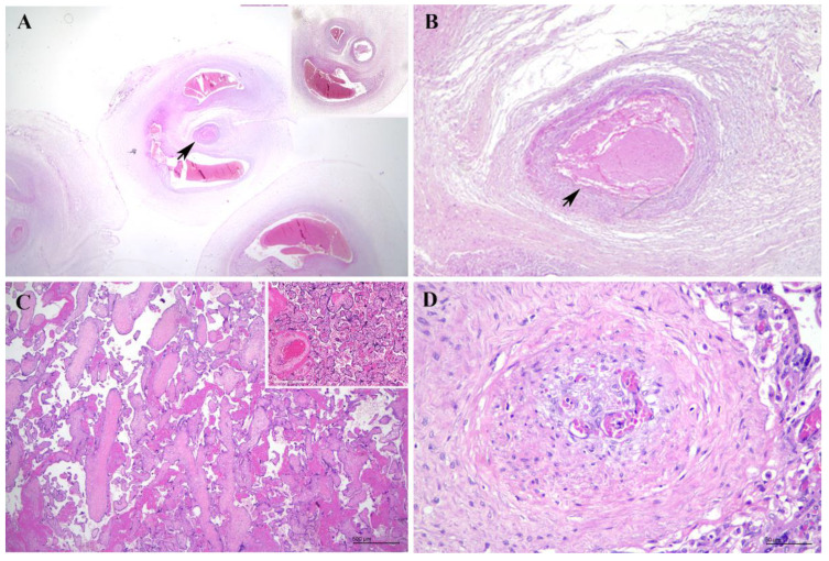
Umbilical artery thrombosis (UAT) masquerading as a single umbilical artery (SUA) is a rare but critical diagnostic challenge in prenatal care. We described a case of a 22-year-old primigravida with an uneventful obstetric history who presented with reduced fetal movements at 22 weeks of gestation. Ultrasound showed no gross fetal structural anomalies while umbilical artery Doppler flow imaging revealed an isolated SUA. The patient again presented with diminished fetal movement at 24 weeks gestation, and a diagnosis of intrauterine demise was confirmed ultrasonographically. She was then induced and delivered a macerated stillborn female fetus. Placental examination revealed three umbilical vessels with an occlusive thrombus seen within the umbilical artery consistent with UAT, a finding previously mistaken for SUA. This case underscores the diagnostic difficulties of UAT radiologically, especially when there was no prior documented evidence of two umbilical arteries. Identification of at-risk fetuses would allow for close monitoring or effective interventions to be implemented as early as possible to avert preventable fetal loss.
伪装成单脐动脉(SUA)的脐动脉血栓形成(UAT)在产前护理中是一种罕见但关键的诊断挑战。我们描述了一例22岁初产妇的病例,其产科病史无异常,在妊娠22周时出现胎动减少。超声检查未发现明显的胎儿结构异常,而脐动脉多普勒血流成像显示为孤立的单脐动脉。该患者在妊娠24周时再次出现胎动减少,超声检查确诊为宫内死亡。随后她接受引产,娩出一个浸软的死产女胎。胎盘检查发现有三条脐血管,在脐动脉内可见闭塞性血栓,符合脐动脉血栓形成,这一发现之前被误诊为单脐动脉。该病例强调了脐动脉血栓形成在影像学上的诊断困难,尤其是在之前没有记录到两条脐动脉的证据时。识别高危胎儿将有助于尽早进行密切监测或实施有效干预,以避免可预防的胎儿丢失。