Department of Cell Biology, Faculty of Biochemistry, Biophysics and Biotechnology, Jagiellonian University, Krakow, Poland.
INSERM U1063, Oxidative Stress and Metabolic Pathologies, Angers University, Angers, France.
J Nanobiotechnology. 2024 Feb 12;22(1):60. doi: 10.1186/s12951-024-02304-y.
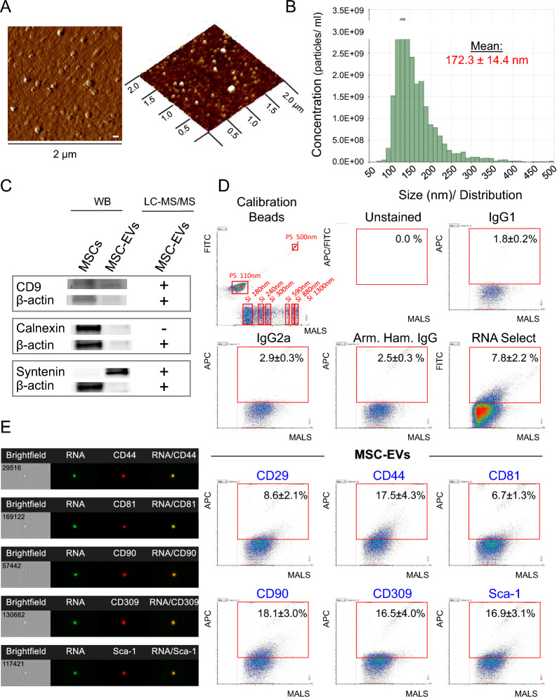
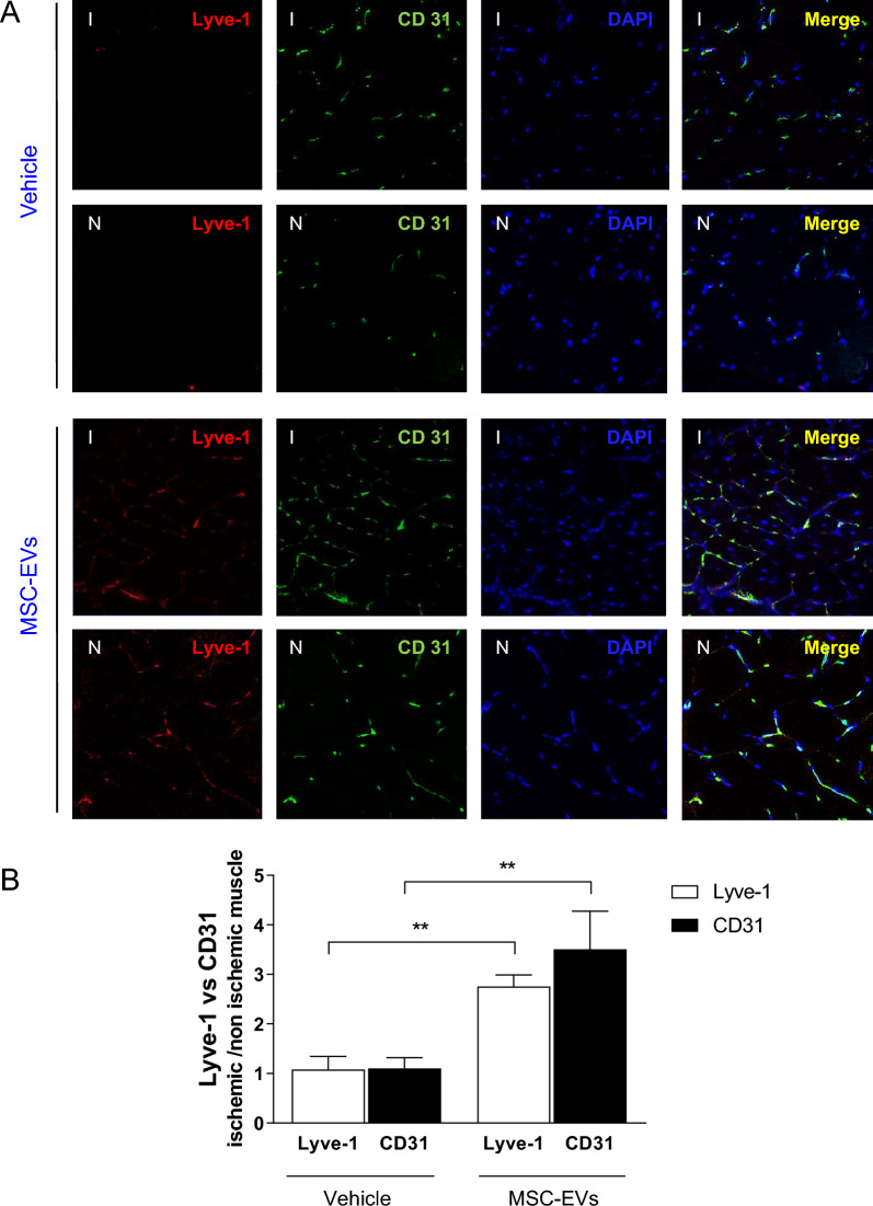
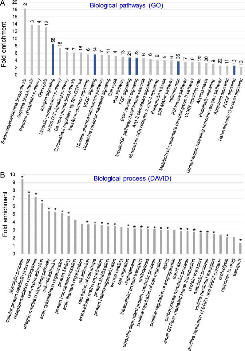
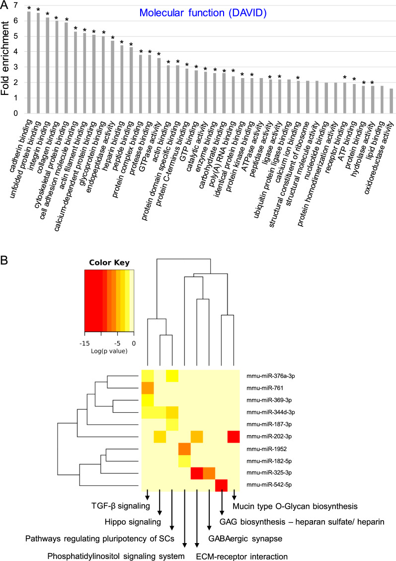
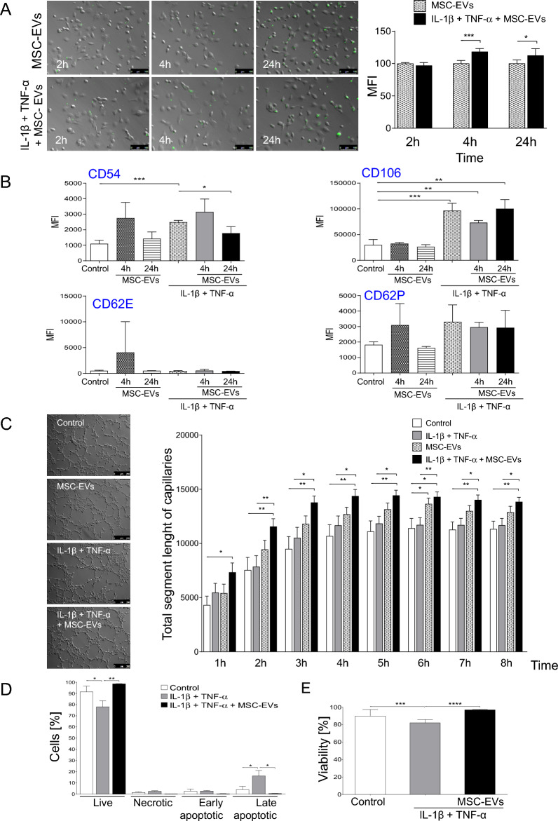
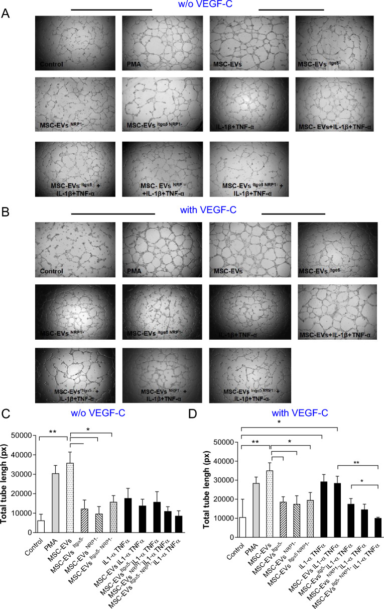
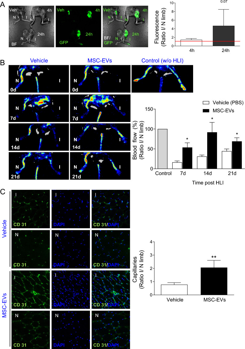
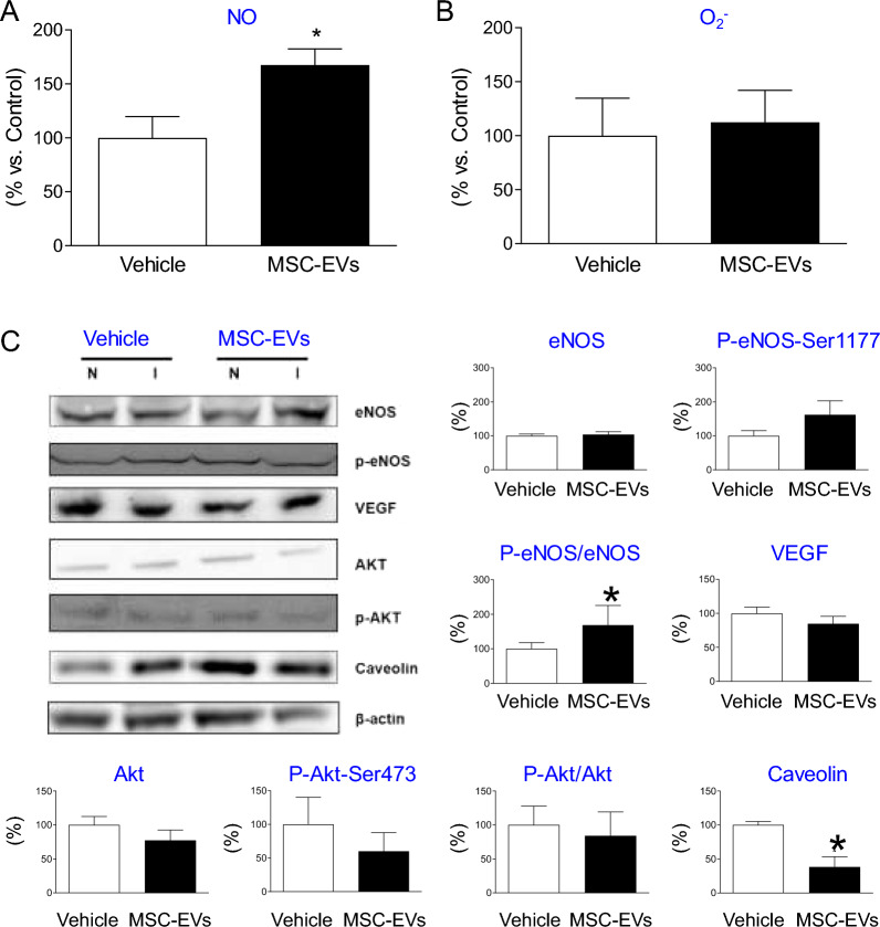
Mesenchymal stem cells/stromal cells (MSCs)-derived extracellular vesicles (EVs) mediate pro-regenerative effects in damaged ischemic tissues by regulating angiogenesis. MSCs-EVs modulate functions of cells including endogenous mature cells, progenitors and stem cells, resulting in restoration of blood flow. However, the mechanisms underlying such MSC-EV activity still remain poorly understood. The present study analyzes biological effects of bone marrow (BM) MSC-EVs on endothelial cells (ECs) in ischemic tissues both in in vitro and in vivo conditions and elucidates the molecular mechanisms underlying the tissue repair. MSC-EVs were isolated from murine BM-derived MSCs and their morphological, antigenic and molecular composition regarding protein and microRNA levels were evaluated to examine their properties. Global proteomic analysis demonstrated the presence in MSC-EVs of proteins regulating pro-regenerative pathways, including integrin α5 (Itgα5) and neuropilin-1 (NRP1) involved in lymphangiogenesis. MSC-EVs were also enriched in microRNAs regulating angiogenesis, TGF-β signaling and processes guiding cellular adhesion and interactions with extracellular matrix. The functional effects of MSC-EVs on capillary ECs in vitro included the increase of capillary-like tube formation and cytoprotection under normal and inflammatory conditions by inhibiting apoptosis. Notably, MSC-EVs enhanced also capillary-like tube formation of lymphatic ECs, which may be regulated by Itgα5 and NRP1. Moreover, in a mouse model of critical hind limb ischemia, MSC-EVs increased the recovery of blood flow in ischemic muscle tissue, which was accompanied with increased vascular density in vivo. This pro-angiogenic effect was associated with an increase in nitric oxide (NO) production via endothelial NO-synthase activation in ischemic muscles. Interestingly, MSC-EVs enhanced lymphangiogenesis, which has never been reported before. The study provides evidence on pro-angiogenic and novel pro-lymphangiogenic role of MSC-EVs on ECs in ischemic tissue mediated by their protein and miRNA molecular cargos. The results highlight Itgα5 and NRP1 carried by MSC-EVs as potential therapeutic targets to boost lymphangiogenesis.
间充质干细胞/基质细胞 (MSC) 衍生的细胞外囊泡 (EV) 通过调节血管生成来介导受损缺血组织中的促再生作用。MSC-EVs 调节包括内源性成熟细胞、祖细胞和干细胞在内的细胞的功能,从而恢复血流。然而,这种 MSC-EV 活性的机制仍知之甚少。本研究分析了骨髓 (BM) MSC-EVs 在体外和体内条件下对缺血组织内皮细胞 (EC) 的生物学作用,并阐明了组织修复的分子机制。从鼠源性 BM 衍生 MSC 中分离 MSC-EVs,评估其形态、抗原和分子组成,包括参与淋巴管生成的整合素 α5 (Itgα5) 和神经纤毛蛋白-1 (NRP1) 的蛋白和 microRNA 水平,以检查其特性。全局蛋白质组学分析表明,MSC-EVs 中存在调节促再生途径的蛋白质,包括参与淋巴管生成的整合素 α5 (Itgα5) 和神经纤毛蛋白-1 (NRP1)。MSC-EVs 还富含调节血管生成、TGF-β 信号转导和指导细胞黏附和与细胞外基质相互作用的过程的 microRNAs。MSC-EVs 对体外毛细血管 EC 的功能影响包括在正常和炎症条件下增加毛细血管样管形成和细胞保护,通过抑制细胞凋亡。值得注意的是,MSC-EVs 还增强了淋巴管内皮细胞的毛细血管样管形成,这可能受到 Itgα5 和 NRP1 的调节。此外,在严重下肢缺血的小鼠模型中,MSC-EVs 增加了缺血肌肉组织的血流恢复,同时体内血管密度增加。这种促血管生成作用与缺血肌肉中内皮型一氧化氮合酶激活导致一氧化氮 (NO) 产生增加有关。有趣的是,MSC-EVs 增强了淋巴管生成,这是以前从未报道过的。该研究提供了证据,证明 MSC-EVs 通过其蛋白质和 miRNA 分子 cargos 对缺血组织中的 EC 具有促血管生成和新型促淋巴管生成作用。研究结果突出了 MSC-EVs 携带的 Itgα5 和 NRP1 作为增强淋巴管生成的潜在治疗靶点。