Department of Neurology, Second Xiangya Hospital, Central South University, Changsha, 410011, People's Republic of China.
The National & Local Joint Engineering Laboratory of Animal Peptide Drug Development, College of Life Sciences, Hunan Normal University, Changsha, 410081, People's Republic of China.
J Nanobiotechnology. 2021 Nov 21;19(1):380. doi: 10.1186/s12951-021-01126-6.
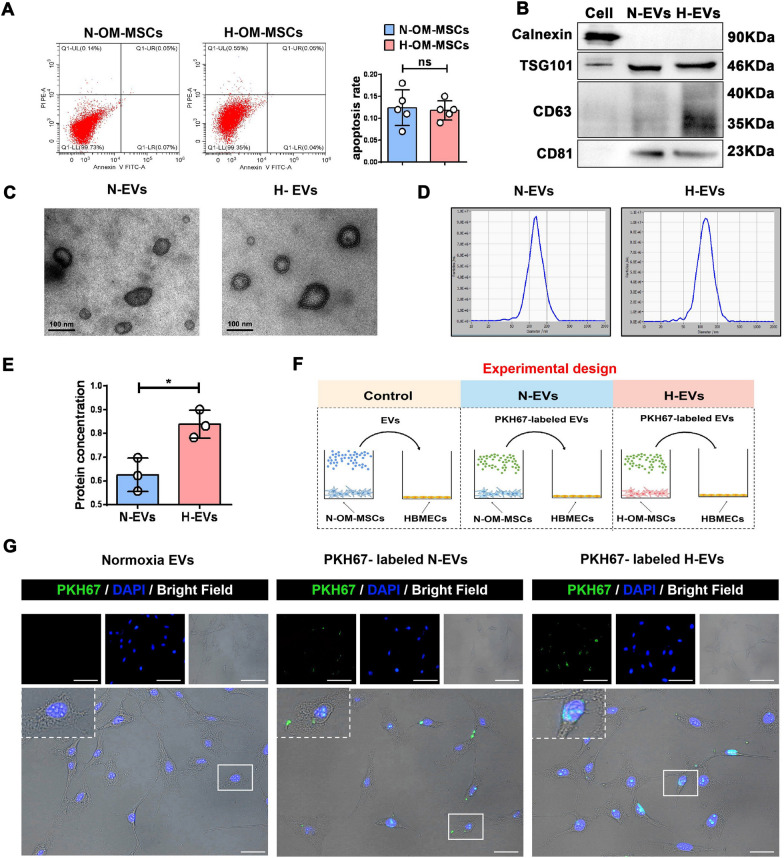
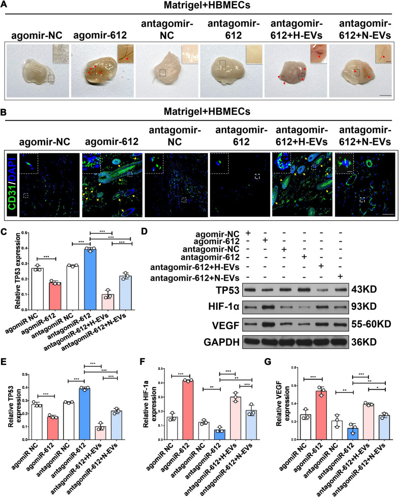
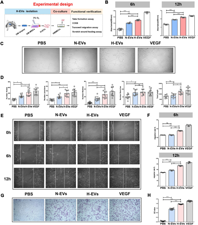
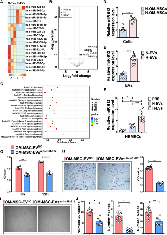
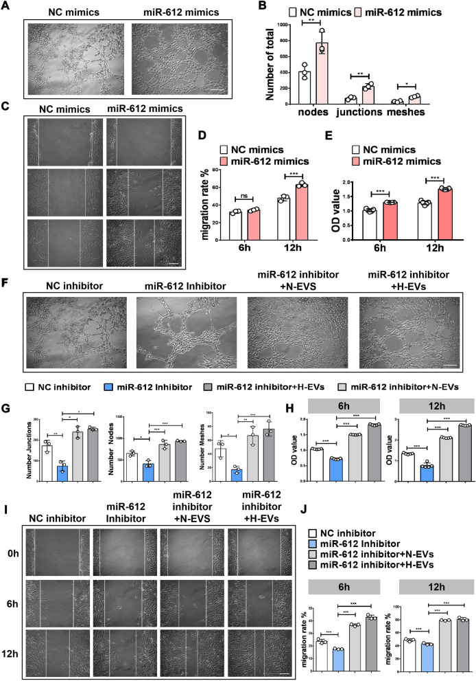
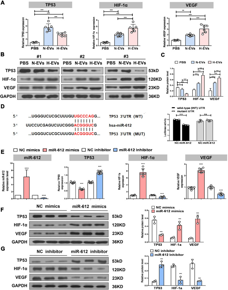
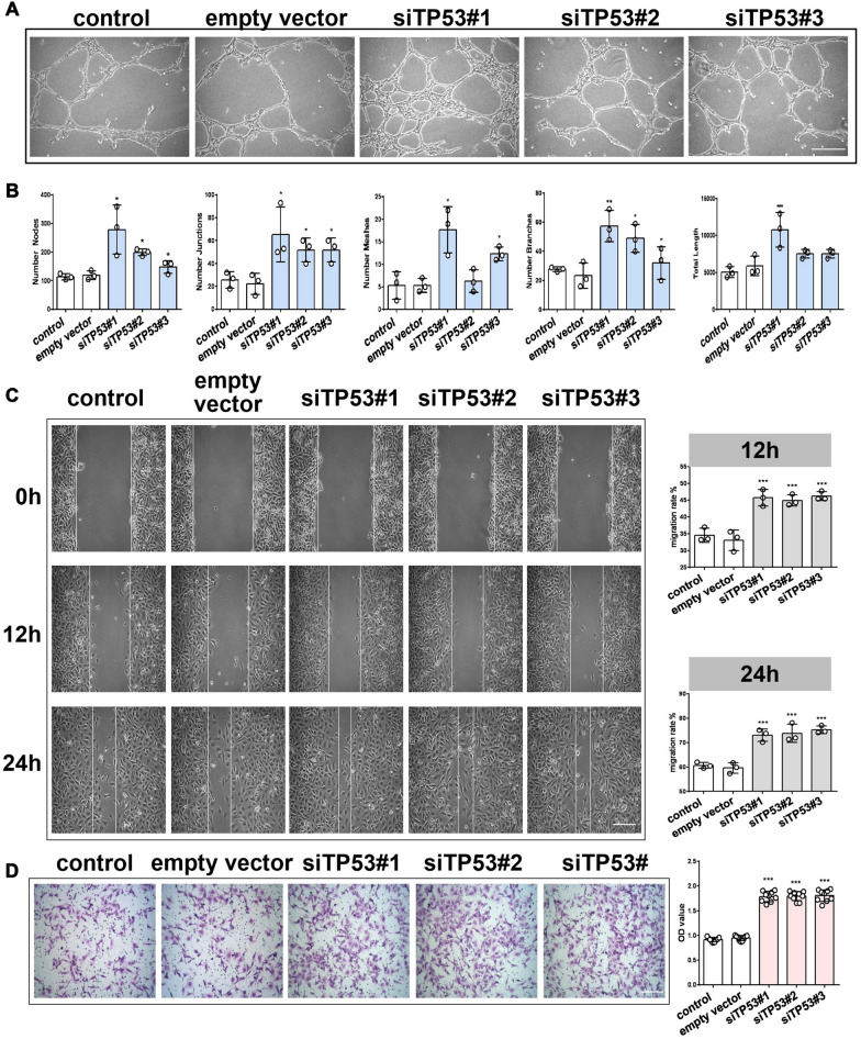
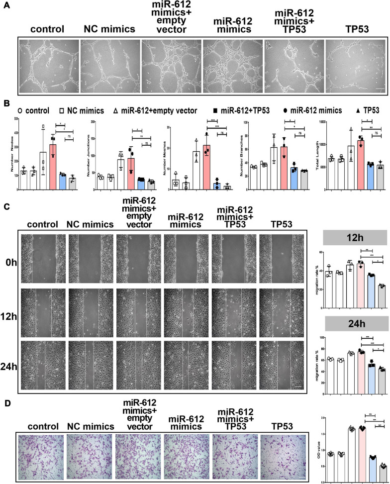
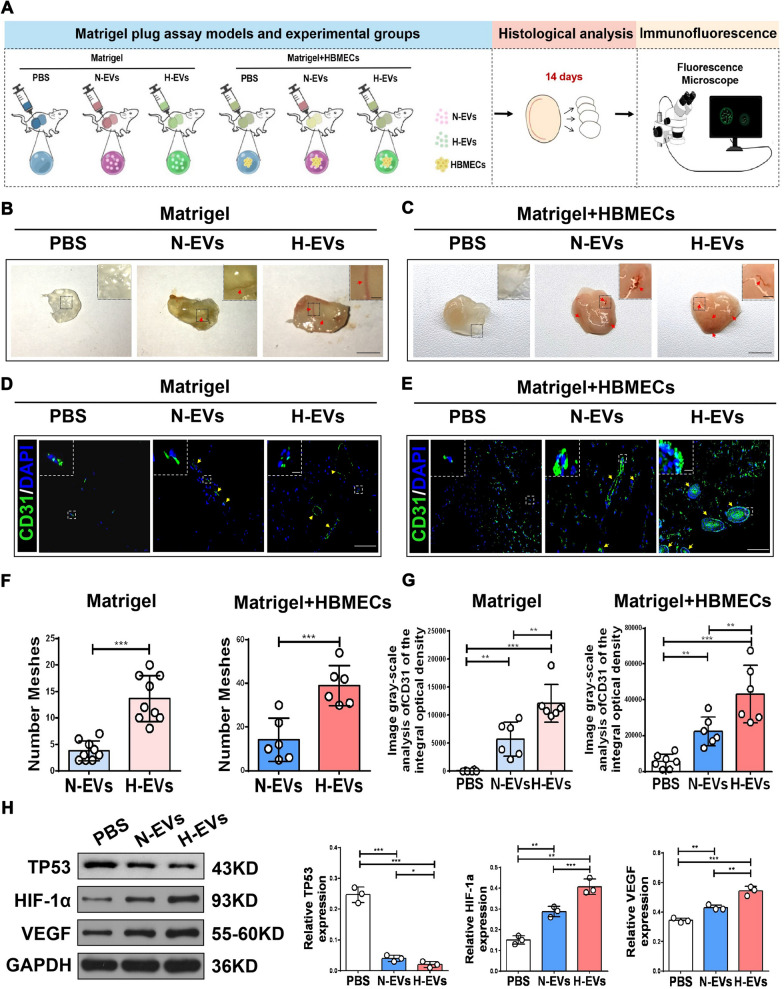
Mesenchymal stem cells (MSCs) play important roles in tissue repair and regeneration, such as the induction of angiogenesis, particularly under hypoxic conditions. However, the molecular mechanisms underlying hypoxic MSC activation remain largely unknown. MSC-derived extracellular vesicles (EVs) are vital mediators of cell-to-cell communication and can be directly utilized as therapeutic agents for tissue repair and regeneration. Here, we explored the effects of EVs from human hypoxic olfactory mucosa MSCs (OM-MSCs) on angiogenesis and its underlying mechanism. EVs were isolated from normoxic (N) OM-MSCs (N-EVs) and hypoxic (H) OM-MSCs (H-EVs) using differential centrifugation and identified by transmission electron microscopy and flow cytometry. In vitro and in vivo, both types of OM-MSC-EVs promoted the proliferation, migration, and angiogenic activities of human brain microvascular endothelial cells (HBMECs). In addition, angiogenesis-stimulatory activity in the H-EV group was significantly enhanced compared to the N-EV group. MicroRNA profiling revealed a higher abundance of miR-612 in H-EVs than in N-EVs, while miR-612 inactivation abolished the N-EV treatment benefit. To explore the roles of miR-612, overexpression and knock-down experiments were performed using a mimic and inhibitor or agomir and antagomir of miR-612. The miR-612 target genes were confirmed using the luciferase reporter assay. Gain- and loss-of-function studies allowed the validation of miR-612 (enriched in hypoxic OM-MSC-EVs) as a functional messenger that stimulates angiogenesis and represses the expression of TP53 by targeting its 3'-untranslated region. Further functional assays showed that hypoxic OM-MSC-EVs promote paracrine Hypoxia-inducible factor 1-alpha (HIF-1α)-Vascular endothelial growth factor (VEGF) signaling in HBMECs via the exosomal miR-612-TP53-HIF-1α-VEGF axis. These findings suggest that hypoxic OM-MSC-EVs may represent a promising strategy for ischemic disease by promoting angiogenesis via miR-612 transfer.
间充质干细胞 (MSCs) 在组织修复和再生中发挥重要作用,例如诱导血管生成,特别是在缺氧条件下。然而,缺氧 MSC 激活的分子机制在很大程度上尚不清楚。MSC 衍生的细胞外囊泡 (EVs) 是细胞间通讯的重要介质,可直接用作组织修复和再生的治疗剂。在这里,我们探讨了缺氧嗅黏膜 MSC(OM-MSCs)衍生的 EVs 对血管生成及其潜在机制的影响。EVs 通过差速离心从常氧(N)OM-MSCs(N-EVs)和缺氧(H)OM-MSCs(H-EVs)中分离出来,并通过透射电子显微镜和流式细胞术进行鉴定。在体外和体内,两种类型的 OM-MSC-EVs 均促进人脑微血管内皮细胞(HBMECs)的增殖、迁移和血管生成活性。此外,与 N-EV 组相比,H-EV 组的血管生成刺激活性显著增强。miRNA 谱分析显示 H-EVs 中 miR-612 的丰度明显高于 N-EVs,而 miR-612 失活消除了 N-EV 处理的益处。为了探讨 miR-612 的作用,使用 miR-612 的模拟物和抑制剂或激动剂和拮抗剂进行了过表达和敲低实验。通过荧光素酶报告基因测定证实了 miR-612 的靶基因。通过增益和失能研究验证了 miR-612(富含缺氧 OM-MSC-EVs)作为一种功能性信使,通过靶向其 3'非翻译区来刺激血管生成并抑制 TP53 的表达。进一步的功能测定表明,缺氧 OM-MSC-EVs 通过外泌体 miR-612-TP53-HIF-1α-VEGF 轴促进 HBMECs 中的 HIF-1α-VEGF 信号传导。这些发现表明,缺氧 OM-MSC-EVs 可能通过转移 miR-612 来促进血管生成,从而成为缺血性疾病的一种很有前途的策略。