Department of Obstetrics and Gynaecology, School of Clinical Medicine, Li Ka Shing Faculty of Medicine, The University of Hong Kong, Pokfulam, China.
Centre for Translational Stem Cell Biology, The University of Hong Kong, Pokfulam, China.
Biol Res. 2024 Feb 13;57(1):6. doi: 10.1186/s40659-024-00484-3.
The monthly regeneration of human endometrial tissue is maintained by the presence of human endometrial mesenchymal stromal/stem cells (eMSC), a cell population co-expressing the perivascular markers CD140b and CD146. Endometrial regeneration is impaired in the presence of intrauterine adhesions, leading to infertility, recurrent pregnancy loss and placental abnormalities. Several types of somatic stem cells have been used to repair the damaged endometrium in animal models, reporting successful pregnancy. However, the ability of endometrial stem cells to repair the damaged endometrium remains unknown.
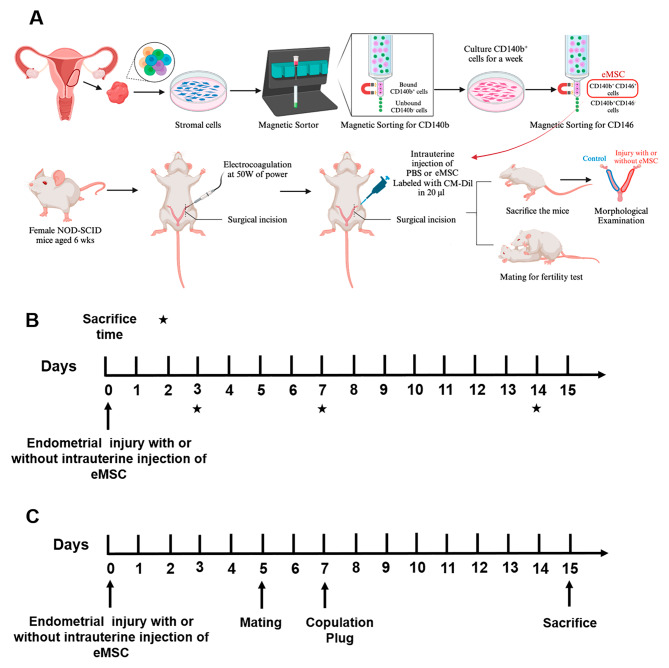
Electrocoagulation was applied to the left uterine horn of NOD/SCID mice causing endometrial injury. Human eMSC or PBS was then injected into the left injured horn while the right normal horn served as controls. Mice were sacrificed at different timepoints (Day 3, 7 and 14) and the endometrial morphological changes as well as the degree of endometrial injury and repair were observed by histological staining. Gene expression of various inflammatory markers was assessed using qPCR. The functionality of the repaired endometrium was evaluated by fertility test.
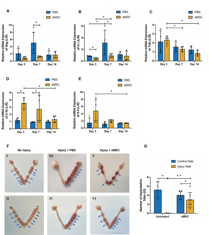
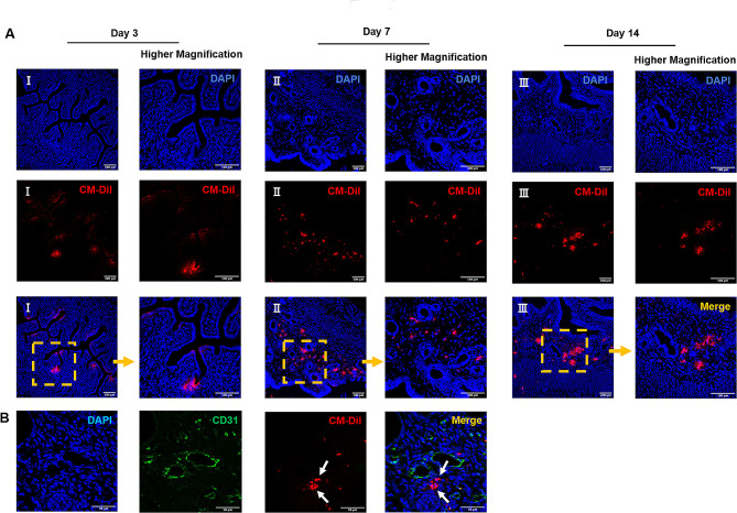
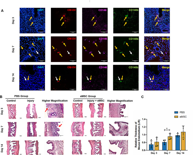
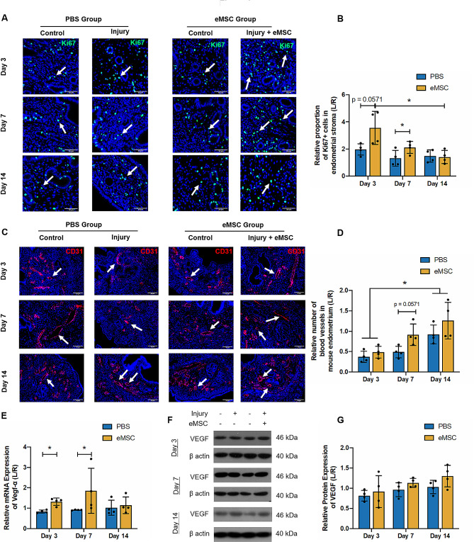
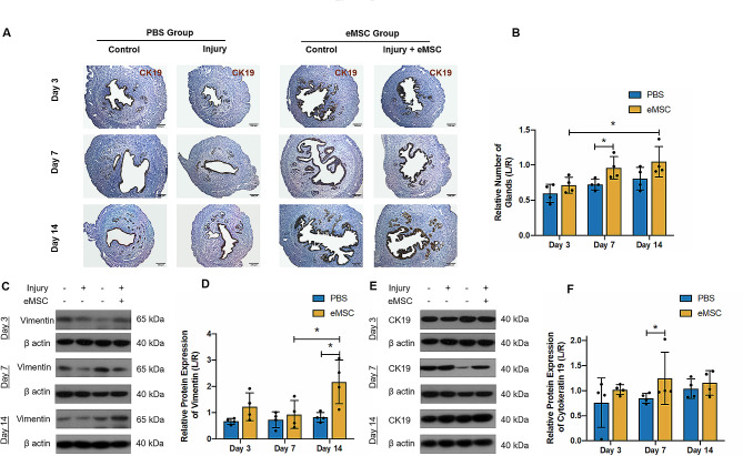
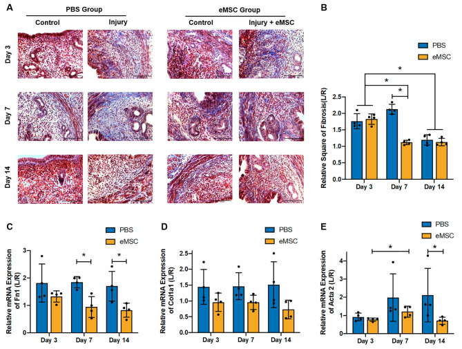
Human eMSC successfully incorporated into the injured uterine horn, which displayed significant morphological restoration. Also, endometrium in the eMSC group showed better cell proliferation and glands formation than the PBS group. Although the number of blood vessels were similar between the two groups, gene expression of VEGF-α significantly increased in the eMSC group. Moreover, eMSC had a positive impact on the regeneration of both stromal and epithelial components of the mouse endometrium, indicated by significantly higher vimentin and CK19 protein expression. Reduced endometrial fibrosis and down-regulation of fibrosis markers were also observed in the eMSC group. The eMSC group had a significantly higher gene expression of anti-inflammatory factor Il-10 and lower mRNA level of pro-inflammatory factors Ifng and Il-2, indicating the role of eMSC in regulation of inflammatory reactions. The eMSC group showed higher implantation sites than the PBS group, suggesting better endometrial receptivity with the presence of newly emerged endometrial lining.
Our findings suggest eMSC improves regeneration of injured endometrium in mice.
人类子宫内膜间质/干基质细胞(eMSC)的存在维持了人类子宫内膜组织的每月再生,该细胞群体共同表达血管周标志物 CD140b 和 CD146。在存在宫腔粘连的情况下,子宫内膜再生受损,导致不孕、反复妊娠丢失和胎盘异常。已经使用几种类型的体干细胞来修复动物模型中的受损子宫内膜,报告了成功妊娠。然而,子宫内膜干细胞修复受损子宫内膜的能力仍不清楚。
用电凝法对 NOD/SCID 小鼠的左侧子宫角造成子宫内膜损伤。然后将人 eMSC 或 PBS 注入左侧受伤的角,而右侧正常角作为对照。在不同时间点(第 3、7 和 14 天)处死小鼠,通过组织学染色观察子宫内膜形态变化以及子宫内膜损伤和修复的程度。使用 qPCR 评估各种炎症标志物的基因表达。通过生育能力测试评估修复后子宫内膜的功能。
人 eMSC 成功整合到受伤的子宫角,显示出明显的形态恢复。此外,eMSC 组的子宫内膜显示出比 PBS 组更好的细胞增殖和腺体形成。尽管两组的血管数量相似,但 eMSC 组中 VEGF-α 的基因表达显著增加。此外,eMSC 对小鼠子宫内膜基质和上皮成分的再生有积极影响,表现为波形蛋白和 CK19 蛋白表达明显升高。eMSC 组还观察到子宫内膜纤维化减少和纤维化标志物下调。eMSC 组抗炎因子 Il-10 的基因表达显著升高,促炎因子 Ifng 和 Il-2 的 mRNA 水平降低,表明 eMSC 在调节炎症反应中的作用。eMSC 组的种植部位明显高于 PBS 组,表明存在新出现的子宫内膜衬里时,子宫内膜接受性更好。
我们的研究结果表明 eMSC 可改善小鼠受伤子宫内膜的再生。