Pathology Department, Medical Faculty of University of Porto, Porto, Portugal.
Pathology Laboratory, Institute of Molecular Pathology and Immunology of University of Porto (IPATIMUP), Rua Júlio Amaral de Carvalho 45, 4200-135, Porto, Portugal.
Virchows Arch. 2024 Jul;485(1):75-82. doi: 10.1007/s00428-024-03762-3. Epub 2024 Feb 14.
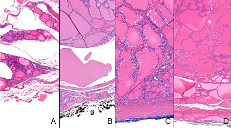
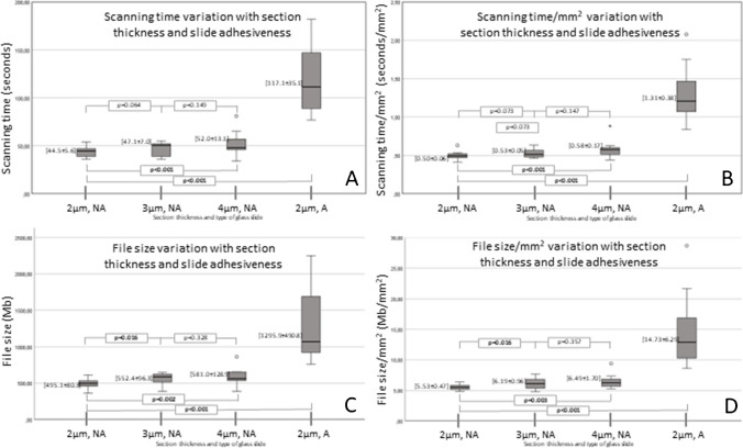
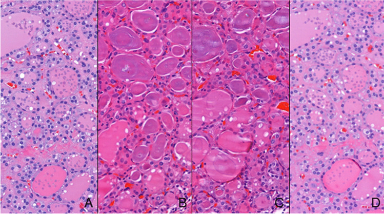
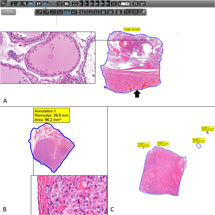
Transition from optical to digital observation requires an additional procedure in the pathology laboratory, the scanning of glass slides, leading to increased time and digital archive consumption. Thyroid surgical samples often carry the need to collect several tissue fragments that generate many slides to be scanned. This study evaluated the impact of using different inking colours for the surgical margin, section thickness, and glass slide type, in the consumption of time and archive. The series comprehended 40 nodules from 30 patients, including 34 benign nodules in follicular nodular disease, 1 NIFTP, and 5 papillary carcinomas. In 12 nodules, the dominant pattern was microfollicular/solid and in 28 it was macrofollicular. Scanning times/mm were longer in red-inked fragments in comparison to green (p = 0.04) and black ones (p = 0.024), and in blue-inked in comparison to green ones (p = 0.043). File sizes/mm were larger in red-inked fragments in comparison to green (p = 0.008) and black ones (p = 0.002). The dominant pattern microfollicular/solid was associated with bigger file size/mm in comparison with the macrofollicular one (p < 0.001). All scanner outputs increase significantly with the thickness of the section. All scanning outputs increase with the usage of adhesive glass slides in comparison to non-adhesive ones. Small interventions in thyroid sample management that can help optimizing the digital workflow include to prefer black and green inking colours for the surgical margins and 2 µm section in non-adhesive glass slides for increased efficiency.
从光学观察向数字观察转变需要在病理实验室进行额外的操作,即扫描载玻片,这会导致时间延长和数字存档消耗增加。甲状腺外科样本通常需要收集多个组织碎片,从而生成许多需要扫描的载玻片。本研究评估了在手术切缘、切片厚度和载玻片类型上使用不同的染色颜色对时间和存档的影响。该系列包括 30 名患者的 40 个结节,其中 34 个为滤泡状结节性疾病的良性结节、1 个非典型滤泡性肿瘤/不能分类的肿瘤、5 个甲状腺乳头状癌。在 12 个结节中,主要模式为微滤泡/实性,在 28 个结节中为大滤泡性。与绿色(p = 0.04)和黑色(p = 0.024)相比,红色墨水标记的碎片扫描时间/毫米更长,与蓝色墨水相比,绿色墨水标记的碎片扫描时间/毫米更长(p = 0.043)。与绿色(p = 0.008)和黑色(p = 0.002)相比,红色墨水标记的碎片文件大小/毫米更大。与大滤泡性相比,微滤泡/实性为主的模式与更大的文件大小/毫米相关(p < 0.001)。所有扫描仪输出都随切片厚度显著增加。与非粘性载玻片相比,使用粘性载玻片会使所有扫描输出增加。在甲状腺样本管理方面进行小的干预可以帮助优化数字工作流程,包括优先选择黑色和绿色墨水标记手术切缘,以及在非粘性载玻片上使用 2μm 切片,以提高效率。