Department of Surgery, Leiden University Medical Center, Leiden, The Netherlands.
Department of Pathology, Leiden University Medical Center, Leiden, The Netherlands.
Sci Rep. 2024 Feb 17;14(1):3983. doi: 10.1038/s41598-024-54718-1.
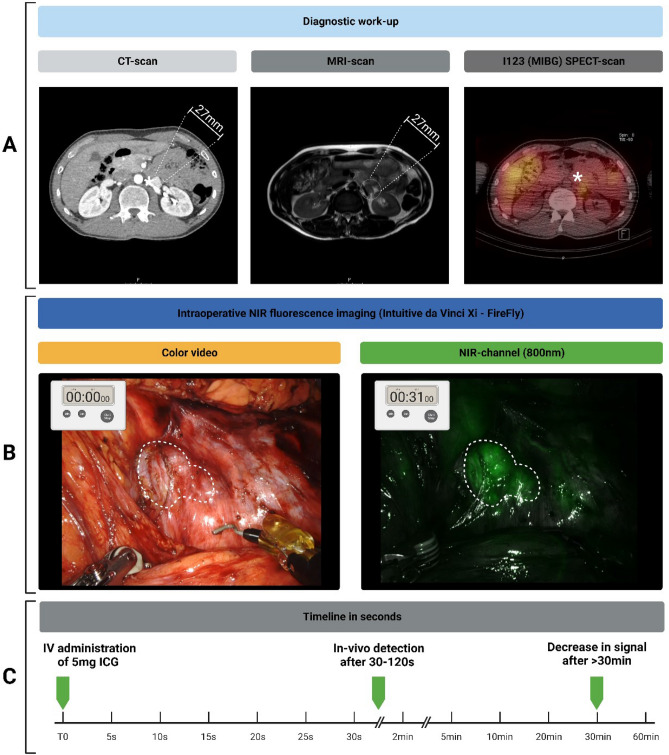
This retrospective study explores the utility of near-infrared (NIR) fluorescence imaging with indocyanine green (ICG) in enhancing the intraoperative identification and guidance for the resection of abdominal paragangliomas. They can be challenging to detect during minimally invasive surgery, due to their anatomical location, varying size and similar appearance in regard to their surrounding tissue. Patients with suspected abdominal paragangliomas planned for a minimally-invasive resection were included. As part of standard of care they received single intravenous dose of 5 mg ICG after abdominal exploration. NIR fluorescence imaging of the anatomical region of the suspected lesion was performed immediately following intravenous administration, to assess fluorescence signals, intraoperative identification, and histopathological correlation. Out of five resected suspicious lesions, four were imaged with NIR fluorescence, pathology confirming four as paragangliomas, the latter turned out to be an adrenal adenoma. NIR fluorescence identified all four lesions, surpassing the limitations of white-light visualization. Homogeneous fluorescence signals appeared 30-60 s post-ICG administration, which lasted up to 30 min. The study demonstrates the feasibility and potential clinical value of fluorescence-guided minimally-invasive resections of abdominal paragangliomas using a single intravenous ICG dose. These findings support the scientific basis for routine use of ICG-fluorescence-guided surgery in challenging anatomical cases, providing valuable assistance in lesion detection and resection.
本回顾性研究探讨了近红外(NIR)荧光成像联合吲哚菁绿(ICG)在增强腹部嗜铬细胞瘤微创手术中识别和指导切除的作用。由于其解剖位置、大小不一,与周围组织相似,这些肿瘤在微创手术中难以检测。本研究纳入了计划行微创切除术的疑似腹部嗜铬细胞瘤患者。作为标准治疗的一部分,他们在腹部探查后接受了 5mgICG 的单次静脉注射。在静脉注射后立即对疑似病变的解剖区域进行 NIR 荧光成像,以评估荧光信号、术中识别和组织病理学相关性。在切除的五个可疑病变中,有四个进行了 NIR 荧光成像,病理证实四个为嗜铬细胞瘤,其余一个为肾上腺腺瘤。NIR 荧光成功识别了所有四个病变,克服了白光可视化的局限性。ICG 给药后 30-60 秒出现均匀荧光信号,持续 30 分钟。本研究表明,单次静脉注射 ICG 进行 NIR 荧光引导的腹部嗜铬细胞瘤微创手术是可行的,具有潜在的临床价值。这些发现为在具有挑战性的解剖病例中常规使用 ICG 荧光引导手术提供了科学依据,为病变检测和切除提供了有价值的帮助。