Division of Life Science, Graduate School of Science and Engineering, Saitama University, Saitama, Japan.
Department of Aquatic Environment and Resource Management, Sher-e-Bangla Agricultural University, Dhaka, Bangladesh.
Dev Growth Differ. 2024 Apr;66(3):219-234. doi: 10.1111/dgd.12917. Epub 2024 Feb 20.
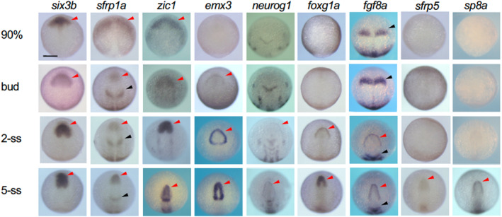
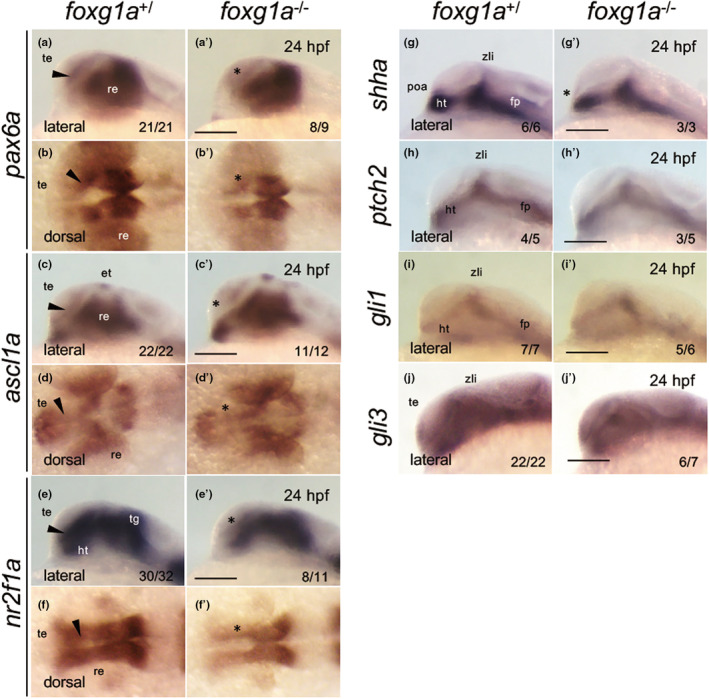
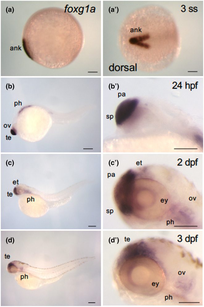
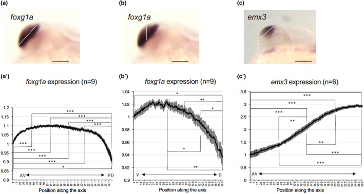
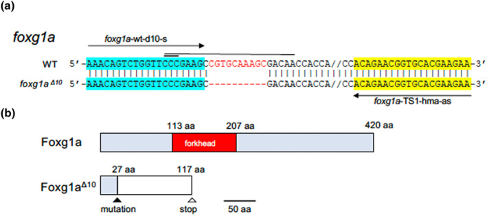
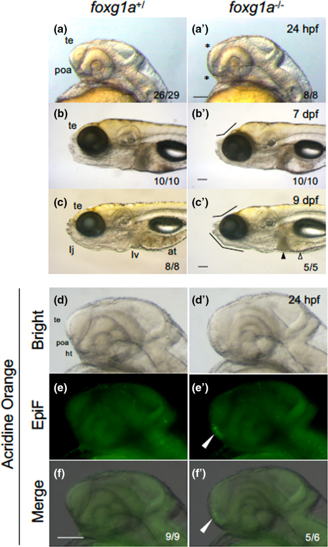
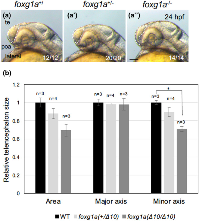
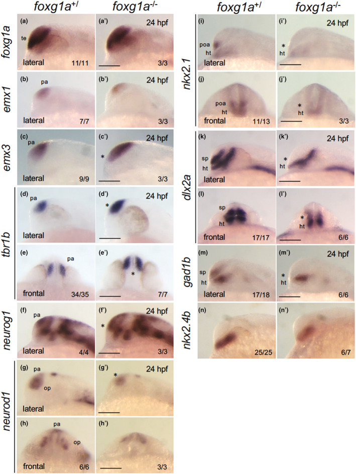
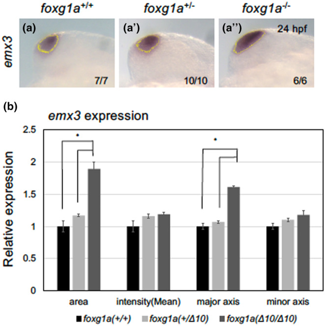
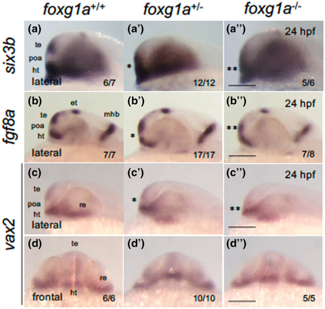
The vertebrate telencephalic lobes consist of the pallium (dorsal) and subpallium (ventral). The subpallium gives rise to the basal ganglia, encompassing the pallidum and striatum. The development of this region is believed to depend on Foxg1/Foxg1a functions in both mice and zebrafish. This study aims to elucidate the genetic regulatory network controlled by foxg1a in subpallium development using zebrafish as a model. The expression gradient of foxg1a within the developing telencephalon was examined semi-quantitatively in initial investigations. Utilizing the CRISPR/Cas9 technique, we subsequently established a foxg1a mutant line and observed the resultant phenotypes. Morphological assessment revealed that foxg1a mutants exhibit a thin telencephalon together with a misshapen preoptic area (POA). Notably, accumulation of apoptotic cells was identified in this region. In mutants at 24 h postfertilization, the expression of pallium markers expanded ventrally, while that of subpallium markers was markedly suppressed. Concurrently, the expression of fgf8a, vax2, and six3b was shifted ventrally, causing anomalous expression in regions typical of POA formation in wild-type embryos. Consequently, the foxg1a mutation led to expansion of the pallium and disrupted the subpallium and POA. This highlights a pivotal role of foxg1a in directing the dorsoventral patterning of the telencephalon, particularly in subpallium differentiation, mirroring observations in mice. Additionally, reduced expression of neural progenitor maintenance genes was detected in mutants, suggesting the necessity of foxg1a in preserving neural progenitors. Collectively, these findings underscore evolutionarily conserved functions of foxg1 in the development of the subpallium in vertebrate embryos.
脊椎动物端脑的脑叶由皮层(背侧)和皮层下(腹侧)组成。皮层下产生基底神经节,包括苍白球和纹状体。这一区域的发育被认为依赖于小鼠和斑马鱼中的 Foxg1/Foxg1a 功能。本研究旨在利用斑马鱼作为模型,阐明 Foxg1a 在皮层下发育过程中控制的遗传调控网络。在初步研究中,对半定量地研究了 foxg1a 在发育中的端脑中的表达梯度。利用 CRISPR/Cas9 技术,我们随后建立了 foxg1a 突变系,并观察了由此产生的表型。形态学评估表明,foxg1a 突变体表现为薄端脑,伴有前脑区(POA)畸形。值得注意的是,该区域发现细胞凋亡细胞堆积。在受精后 24 小时的突变体中,皮层标记物的表达向腹侧扩展,而皮层下标记物的表达则明显受到抑制。同时,fgf8a、vax2 和 six3b 的表达向腹侧转移,导致野生型胚胎中典型 POA 形成区域的异常表达。因此,foxg1a 突变导致了皮层的扩张,并破坏了皮层下和 POA。这表明 foxg1a 在端脑的背腹侧模式形成中起着关键作用,特别是在皮层下分化中,与小鼠的观察结果一致。此外,在突变体中检测到神经祖细胞维持基因的表达减少,表明 foxg1a 对于维持神经祖细胞是必要的。总之,这些发现强调了 foxg1 在脊椎动物胚胎中皮层下发育中的保守作用。