• 文献检索
  • 文档翻译
  • 深度研究
  • 学术资讯
  • Suppr Zotero 插件Zotero 插件
  • 邀请有礼
  • 套餐&价格
  • 历史记录
应用&插件
Suppr Zotero 插件Zotero 插件浏览器插件Mac 客户端Windows 客户端微信小程序
定价
高级版会员购买积分包购买API积分包
服务
文献检索文档翻译深度研究API 文档MCP 服务
关于我们
关于 Suppr公司介绍联系我们用户协议隐私条款
关注我们

Suppr 超能文献

核心技术专利:CN118964589B侵权必究
粤ICP备2023148730 号-1Suppr @ 2026

文献检索

告别复杂PubMed语法,用中文像聊天一样搜索,搜遍4000万医学文献。AI智能推荐,让科研检索更轻松。

立即免费搜索

文件翻译

保留排版,准确专业,支持PDF/Word/PPT等文件格式,支持 12+语言互译。

免费翻译文档

深度研究

AI帮你快速写综述,25分钟生成高质量综述,智能提取关键信息,辅助科研写作。

立即免费体验

血脑屏障功能障碍可预测幼年大鼠创伤性脑损伤后的小胶质细胞激活。

Blood-Brain Barrier Dysfunction Predicts Microglial Activation After Traumatic Brain Injury in Juvenile Rats.

作者信息

Green Tabitha R F, Nguyen Tina, Dunker Veronika, Ashton Danielle, Ortiz J Bryce, Murphy Sean M, Rowe Rachel K

机构信息

Department of Integrative Physiology, University of Colorado Boulder, Colorado, USA.

Department of Child Health, University of Arizona College of Medicine-Phoenix, Arizona, USA.

出版信息

Neurotrauma Rep. 2024 Feb 8;5(1):95-116. doi: 10.1089/neur.2023.0057. eCollection 2024.

DOI:10.1089/neur.2023.0057
PMID:38404523
原文链接:https://pmc.ncbi.nlm.nih.gov/articles/PMC10890961/
Abstract

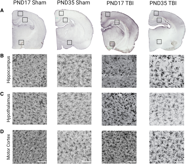
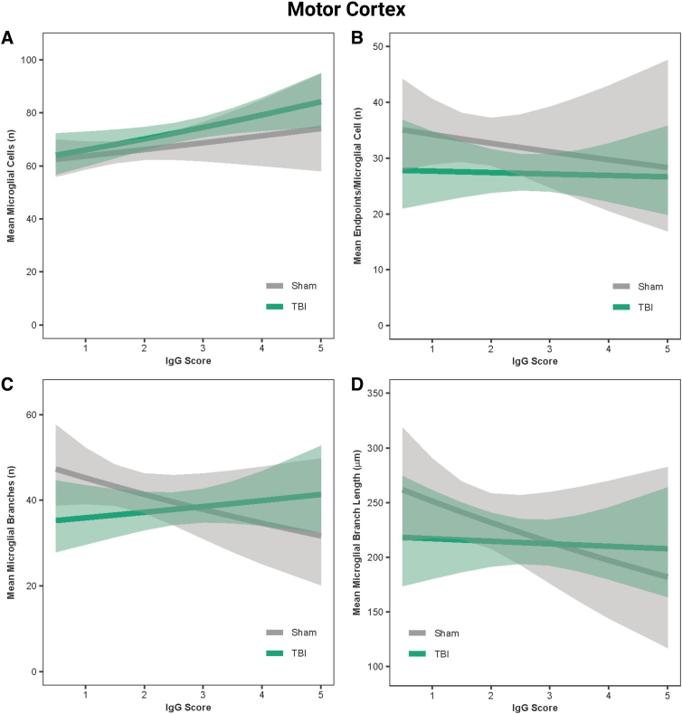
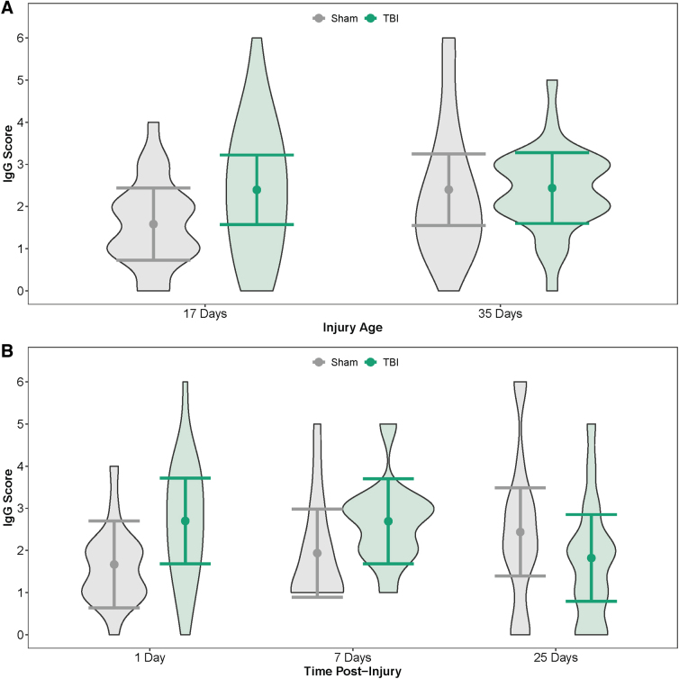
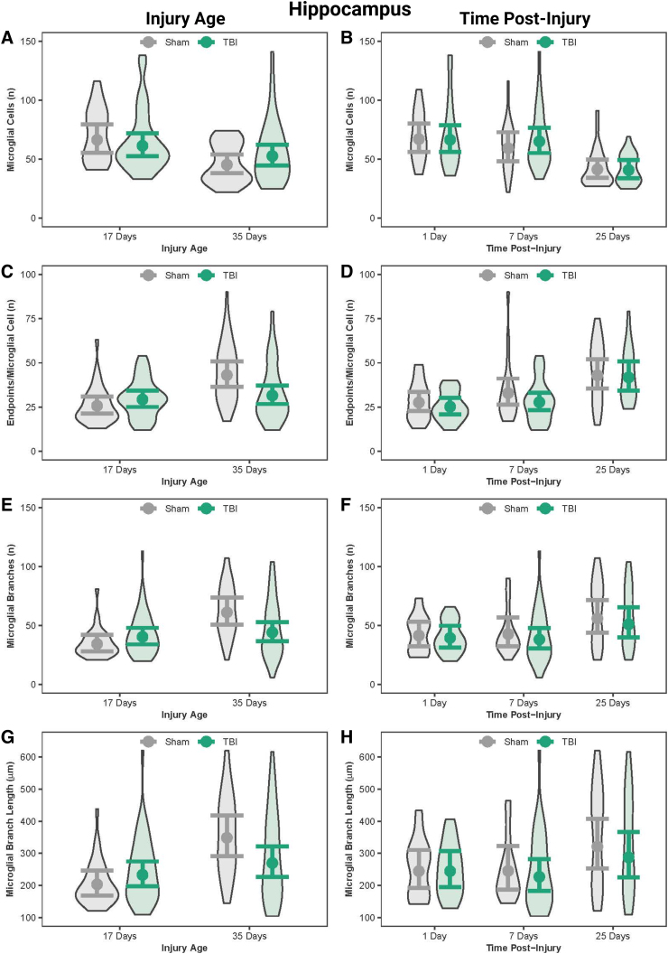
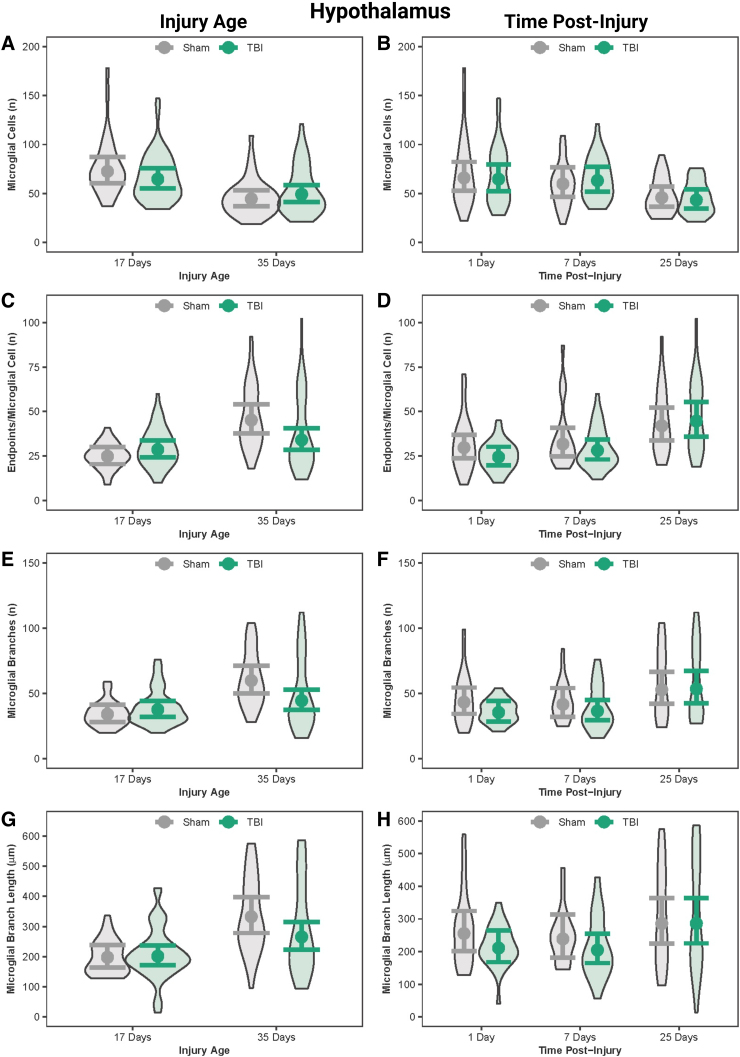
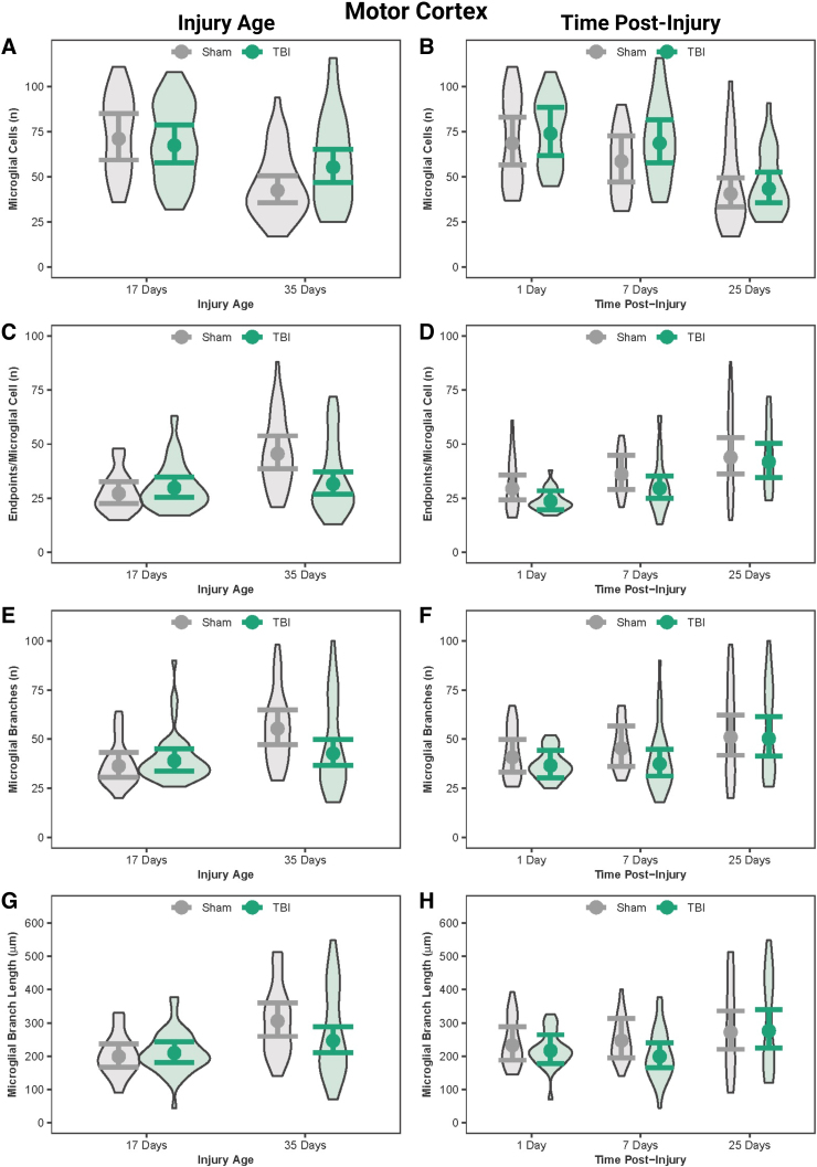
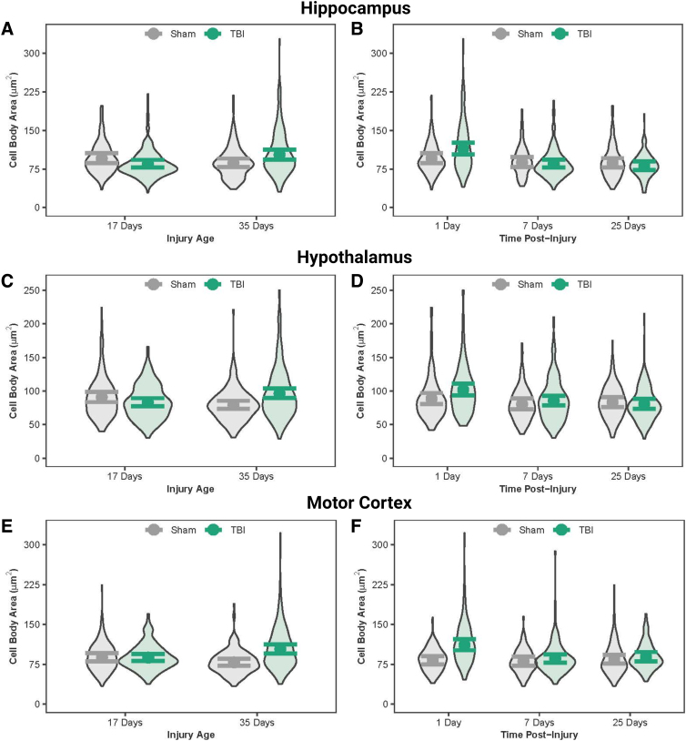
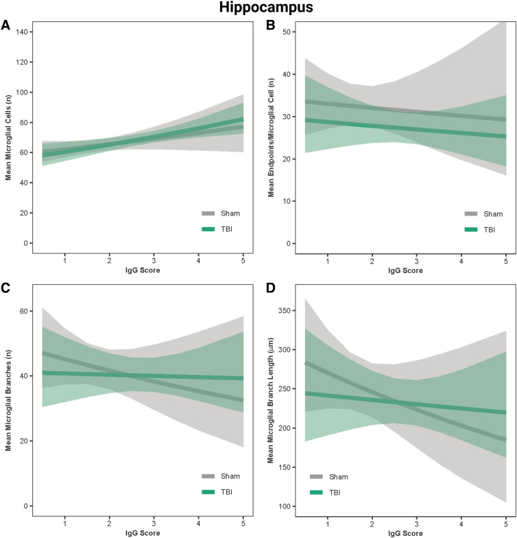
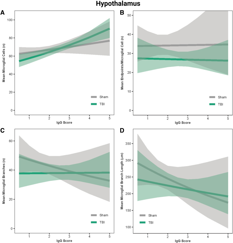
Traumatic brain injury (TBI) disrupts the blood-brain barrier (BBB), which may exacerbate neuroinflammation post-injury. Few translational studies have examined BBB dysfunction and subsequent neuroinflammation post-TBI in juveniles. We hypothesized that BBB dysfunction positively predicts microglial activation and that vulnerability to BBB dysfunction and associated neuroinflammation are dependent on age at injury. Post-natal day (PND)17 and PND35 rats ( = 56) received midline fluid percussion injury or sham surgery, and immunoglobulin-G (IgG) stain was quantified as a marker of extravasated blood in the brain and BBB dysfunction. We investigated BBB dysfunction and the microglial response in the hippocampus, hypothalamus, and motor cortex relative to age at injury and days post-injury (DPI; 1, 7, and 25). We measured the morphologies of ionized calcium-binding adaptor molecule 1-labeled microglia using cell body area and perimeter, microglial branch number and length, end-points/microglial cell, and number of microglia. Data were analyzed using generalized hierarchical models. In PND17 rats, TBI increased levels of IgG compared to shams. Independent of age at injury, IgG in TBI rats was higher at 1 and 7 DPI, but resolved by 25 DPI. TBI activated microglia (more cells and fewer end-points) in PND35 rats compared to respective shams. Independent of age at injury, TBI induced morphological changes indicative of microglial activation, which resolved by 25 DPI. TBI rats had fewer cells and end-points per cell at 1 and 7 DPI than 25 DPI. Independent of TBI, PND17 rats had larger, more activated microglia than PND35 rats; PND17 TBI rats had larger cell body areas and perimeters than PND35 TBI rats. Importantly, we found support in both ages that IgG quantification predicted microglial activation after TBI. The number of microglia increased with increasing IgG, whereas branch length decreased with increasing IgG, which together indicate microglial activation. Our results suggest that stabilization of the BBB after pediatric TBI may be an important therapeutic strategy to limit neuroinflammation and promote recovery.

摘要

创伤性脑损伤(TBI)会破坏血脑屏障(BBB),这可能会加剧损伤后的神经炎症。很少有转化研究探讨青少年TBI后的血脑屏障功能障碍及随后的神经炎症。我们假设血脑屏障功能障碍可正向预测小胶质细胞激活,且血脑屏障功能障碍及相关神经炎症的易感性取决于受伤时的年龄。出生后第17天(PND17)和第35天(PND35)的大鼠(n = 56)接受中线流体冲击伤或假手术,免疫球蛋白G(IgG)染色被定量作为脑内血管外渗血液和血脑屏障功能障碍的标志物。我们研究了相对于受伤时年龄和伤后天数(DPI;1、7和25天),海马体、下丘脑和运动皮层中的血脑屏障功能障碍及小胶质细胞反应。我们使用细胞体面积和周长、小胶质细胞分支数量和长度、端点/小胶质细胞以及小胶质细胞数量来测量离子钙结合衔接分子1标记的小胶质细胞的形态。数据使用广义分层模型进行分析。在PND17大鼠中,与假手术组相比,TBI增加了IgG水平。与受伤时年龄无关,TBI大鼠在1和7 DPI时IgG水平较高,但在25 DPI时恢复正常。与各自的假手术组相比,TBI激活了PND35大鼠中的小胶质细胞(细胞更多且端点更少)。与受伤时年龄无关,TBI诱导了表明小胶质细胞激活的形态学变化,这些变化在25 DPI时消失。TBI大鼠在1和7 DPI时每个细胞的细胞和端点比25 DPI时少。与TBI无关,PND17大鼠的小胶质细胞比PND35大鼠更大且更活跃;PND17 TBI大鼠的细胞体面积和周长比PND35 TBI大鼠更大。重要的是,我们在两个年龄段都发现IgG定量可预测TBI后的小胶质细胞激活。小胶质细胞数量随IgG增加而增加,而分支长度随IgG增加而减少,这共同表明小胶质细胞激活。我们的结果表明,小儿TBI后稳定血脑屏障可能是限制神经炎症和促进恢复的重要治疗策略。

https://cdn.ncbi.nlm.nih.gov/pmc/blobs/b178/10890961/896eec3c9cbc/neur.2023.0057_figure9.jpg
https://cdn.ncbi.nlm.nih.gov/pmc/blobs/b178/10890961/da639138b098/neur.2023.0057_figure1.jpg
https://cdn.ncbi.nlm.nih.gov/pmc/blobs/b178/10890961/66b3ff7ab29f/neur.2023.0057_figure2.jpg
https://cdn.ncbi.nlm.nih.gov/pmc/blobs/b178/10890961/21b81ffa07c4/neur.2023.0057_figure3.jpg
https://cdn.ncbi.nlm.nih.gov/pmc/blobs/b178/10890961/c1dabdfa7bdb/neur.2023.0057_figure4.jpg
https://cdn.ncbi.nlm.nih.gov/pmc/blobs/b178/10890961/fd901867ffd0/neur.2023.0057_figure5.jpg
https://cdn.ncbi.nlm.nih.gov/pmc/blobs/b178/10890961/acf38979713e/neur.2023.0057_figure6.jpg
https://cdn.ncbi.nlm.nih.gov/pmc/blobs/b178/10890961/0b7e8b9f3ad2/neur.2023.0057_figure7.jpg
https://cdn.ncbi.nlm.nih.gov/pmc/blobs/b178/10890961/6c2ab45e71b1/neur.2023.0057_figure8.jpg
https://cdn.ncbi.nlm.nih.gov/pmc/blobs/b178/10890961/896eec3c9cbc/neur.2023.0057_figure9.jpg

相似文献

1
Blood-Brain Barrier Dysfunction Predicts Microglial Activation After Traumatic Brain Injury in Juvenile Rats.血脑屏障功能障碍可预测幼年大鼠创伤性脑损伤后的小胶质细胞激活。
Neurotrauma Rep. 2024 Feb 8;5(1):95-116. doi: 10.1089/neur.2023.0057. eCollection 2024.
2
Age-At-Injury Influences the Glial Response to Traumatic Brain Injury in the Cortex of Male Juvenile Rats.受伤年龄影响雄性幼年大鼠皮层对创伤性脑损伤的胶质细胞反应。
Front Neurol. 2022 Jan 17;12:804139. doi: 10.3389/fneur.2021.804139. eCollection 2021.
3
Midline (central) fluid percussion model of traumatic brain injury in pediatric and adolescent rats.小儿和青少年大鼠创伤性脑损伤的中线(中央)流体冲击模型
J Neurosurg Pediatr. 2018 Jul;22(1):22-30. doi: 10.3171/2018.1.PEDS17449. Epub 2018 Apr 20.
4
Long-lasting blood-brain barrier dysfunction and neuroinflammation after traumatic brain injury.创伤性脑损伤后持久的血脑屏障功能障碍和神经炎症。
Neurobiol Dis. 2020 Nov;145:105080. doi: 10.1016/j.nbd.2020.105080. Epub 2020 Sep 9.
5
Traumatic Brain Injury Causes Chronic Cortical Inflammation and Neuronal Dysfunction Mediated by Microglia.创伤性脑损伤导致慢性皮质炎症和神经元功能障碍,其介导作用是小胶质细胞。
J Neurosci. 2021 Feb 17;41(7):1597-1616. doi: 10.1523/JNEUROSCI.2469-20.2020. Epub 2021 Jan 15.
6
Chronic Cortical Inflammation, Cognitive Impairment, and Immune Reactivity Associated with Diffuse Brain Injury Are Ameliorated by Forced Turnover of Microglia.慢性皮质炎症、认知障碍和弥漫性脑损伤相关的免疫反应可通过小胶质细胞的强制更替得到改善。
J Neurosci. 2022 May 18;42(20):4215-4228. doi: 10.1523/JNEUROSCI.1910-21.2022. Epub 2022 Apr 19.
7
Mossy cell hypertrophy and synaptic changes in the hilus following mild diffuse traumatic brain injury in pigs.猪轻度弥漫性创伤性脑损伤后海洛因区苔藓细胞肥大和突触变化。
J Neuroinflammation. 2020 Jan 31;17(1):44. doi: 10.1186/s12974-020-1720-0.
8
Sleep fragmentation after traumatic brain injury impairs behavior and conveys long-lasting impacts on neuroinflammation.创伤性脑损伤后的睡眠碎片化会损害行为,并对神经炎症产生长期影响。
Brain Behav Immun Health. 2024 May 15;38:100797. doi: 10.1016/j.bbih.2024.100797. eCollection 2024 Jul.
9
Cognitive deficits develop 1month after diffuse brain injury and are exaggerated by microglia-associated reactivity to peripheral immune challenge.认知缺陷在弥漫性脑损伤1个月后出现,并因小胶质细胞对外周免疫挑战的相关反应而加剧。
Brain Behav Immun. 2016 May;54:95-109. doi: 10.1016/j.bbi.2016.01.009. Epub 2016 Jan 14.
10
Colony-Stimulating Factor-1 Receptor Inhibition Transiently Attenuated the Peripheral Immune Response to Experimental Traumatic Brain Injury.集落刺激因子-1受体抑制可短暂减弱对实验性创伤性脑损伤的外周免疫反应。
Neurotrauma Rep. 2023 Apr 28;4(1):284-296. doi: 10.1089/neur.2022.0092. eCollection 2023.

引用本文的文献

1
Microglial depletion and repopulation differentially modulate sleep and inflammation in a mouse model of traumatic brain injury.在创伤性脑损伤小鼠模型中,小胶质细胞耗竭和再填充对睡眠和炎症的调节存在差异。
Neurobiol Sleep Circadian Rhythms. 2025 Feb 28;18(Suppl):100115. doi: 10.1016/j.nbscr.2025.100115. eCollection 2025 May.
2
Repetitive concussions promote microglia-mediated engulfment of presynaptic excitatory input associated with cognitive dysfunction.重复性脑震荡会促进小胶质细胞介导的对与认知功能障碍相关的突触前兴奋性输入的吞噬作用。
Commun Biol. 2025 Feb 28;8(1):335. doi: 10.1038/s42003-025-07729-1.
3
Social deficits mirror delayed cerebrovascular dysfunction after traumatic brain injury.

本文引用的文献

1
Comparisons of quantitative approaches for assessing microglial morphology reveal inconsistencies, ecological fallacy, and a need for standardization.定量方法评估小胶质细胞形态的比较研究揭示了不一致性、生态谬误以及标准化的必要性。
Sci Rep. 2022 Oct 28;12(1):18196. doi: 10.1038/s41598-022-23091-2.
2
Reactive morphology of dividing microglia following kainic acid administration.给予海藻酸后分裂小胶质细胞的反应性形态学
Front Neurosci. 2022 Sep 29;16:972138. doi: 10.3389/fnins.2022.972138. eCollection 2022.
3
The effects of early exercise in traumatic brain-injured rats with changes in motor ability, brain tissue, and biomarkers.
社会缺陷反映了创伤性脑损伤后的脑血管功能延迟障碍。
Acta Neuropathol Commun. 2024 Aug 7;12(1):126. doi: 10.1186/s40478-024-01840-w.
早期运动对运动能力、脑组织和生物标志物改变的创伤性脑损伤大鼠的影响。
BMB Rep. 2022 Oct;55(10):512-517. doi: 10.5483/BMBRep.2022.55.10.097.
4
Automatic hemorrhage segmentation on head CT scan for traumatic brain injury using 3D deep learning model.利用 3D 深度学习模型对头 CT 扫描外伤性脑损伤进行自动出血分割。
Comput Biol Med. 2022 Jul;146:105530. doi: 10.1016/j.compbiomed.2022.105530. Epub 2022 Apr 18.
5
Age-At-Injury Influences the Glial Response to Traumatic Brain Injury in the Cortex of Male Juvenile Rats.受伤年龄影响雄性幼年大鼠皮层对创伤性脑损伤的胶质细胞反应。
Front Neurol. 2022 Jan 17;12:804139. doi: 10.3389/fneur.2021.804139. eCollection 2021.
6
Phillyrin Prevents Neuroinflammation-Induced Blood-Brain Barrier Damage Following Traumatic Brain Injury Altering Microglial Polarization.连翘苷通过改变小胶质细胞极化防止创伤性脑损伤后神经炎症诱导的血脑屏障损伤。
Front Pharmacol. 2021 Oct 20;12:719823. doi: 10.3389/fphar.2021.719823. eCollection 2021.
7
Pathophysiology of Pediatric Traumatic Brain Injury.小儿创伤性脑损伤的病理生理学
Front Neurol. 2021 Jul 15;12:696510. doi: 10.3389/fneur.2021.696510. eCollection 2021.
8
Microglia: A Double-Edged Sword in Intracerebral Hemorrhage From Basic Mechanisms to Clinical Research.小胶质细胞:脑出血的双刃剑——从基础机制到临床研究。
Front Immunol. 2021 May 6;12:675660. doi: 10.3389/fimmu.2021.675660. eCollection 2021.
9
Microglial activation and blood-brain barrier permeability in cerebral small vessel disease.脑小血管病中的小胶质细胞激活和血脑屏障通透性。
Brain. 2021 Jun 22;144(5):1361-1371. doi: 10.1093/brain/awab003.
10
Traumatic Brain Injury Causes Chronic Cortical Inflammation and Neuronal Dysfunction Mediated by Microglia.创伤性脑损伤导致慢性皮质炎症和神经元功能障碍,其介导作用是小胶质细胞。
J Neurosci. 2021 Feb 17;41(7):1597-1616. doi: 10.1523/JNEUROSCI.2469-20.2020. Epub 2021 Jan 15.