Department of Child Health, University of Arizona College of Medicine-Phoenix, Phoenix, AZ, USA.
Department of Integrative Physiology, University of Colorado, 2860 Wilderness Place, Boulder, CO, 80301, USA.
Sci Rep. 2022 Oct 28;12(1):18196. doi: 10.1038/s41598-022-23091-2.
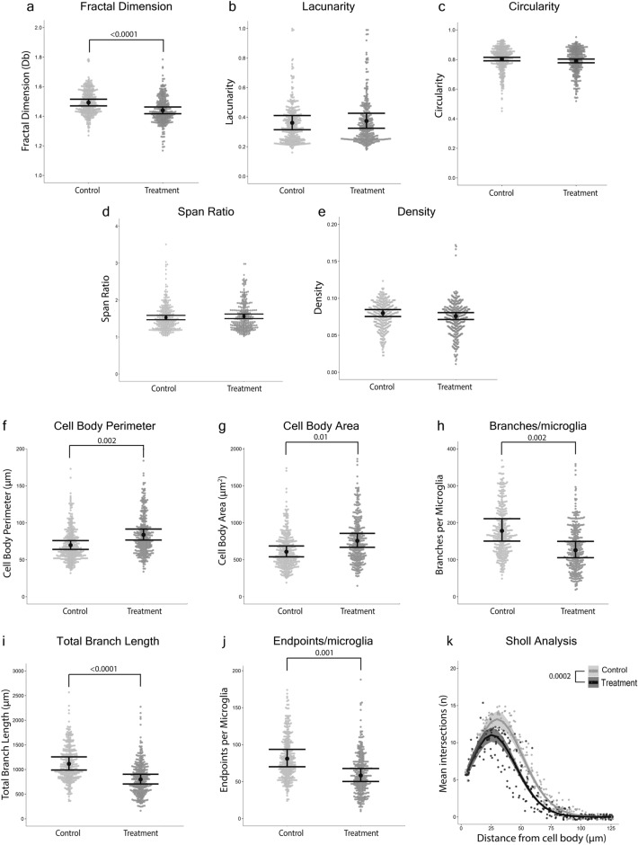
Microglial morphology is used to measure neuroinflammation and pathology. For reliable inference, it is critical that microglial morphology is accurately quantified and that results can be easily interpreted and compared across studies and laboratories. The process through which microglial morphology is quantified is a key methodological choice and little is known about how this choice may bias conclusions. We applied five of the most commonly used ImageJ-based methods for quantifying the microglial morphological response to a stimulus to identical photomicrographs and individual microglial cells isolated from these photomicrographs, which allowed for direct comparisons of results generated using these approaches. We found a lack of comparability across methods that analyzed full photomicrographs, with significant discrepancies in results among the five methods. Quantitative methods to analyze microglial morphology should be selected based on several criteria, and combinations of these methods may give the most biologically accurate representation of microglial morphology.
小胶质细胞形态用于测量神经炎症和病理学。为了进行可靠的推断,准确地量化小胶质细胞形态以及能够轻松地在研究和实验室之间解释和比较结果至关重要。量化小胶质细胞形态的过程是一个关键的方法学选择,而对于这种选择如何影响结论知之甚少。我们应用了五种最常用的基于 ImageJ 的方法来定量分析刺激对小胶质细胞形态的反应,这些方法适用于对来自这些显微照片的单个小胶质细胞进行直接比较。我们发现,对完整显微照片进行分析的方法之间缺乏可比性,五种方法的结果存在显著差异。分析小胶质细胞形态的定量方法应基于几个标准进行选择,并且这些方法的组合可能会最准确地反映小胶质细胞形态的生物学特征。