Biomedical Engineering, Illinois Institute of Technology, Chicago, IL, 60616, USA.
Thayer School of Engineering, Dartmouth College, Hanover, NH, 03755, USA.
Ann Biomed Eng. 2024 Jun;52(6):1625-1637. doi: 10.1007/s10439-024-03476-2. Epub 2024 Feb 26.
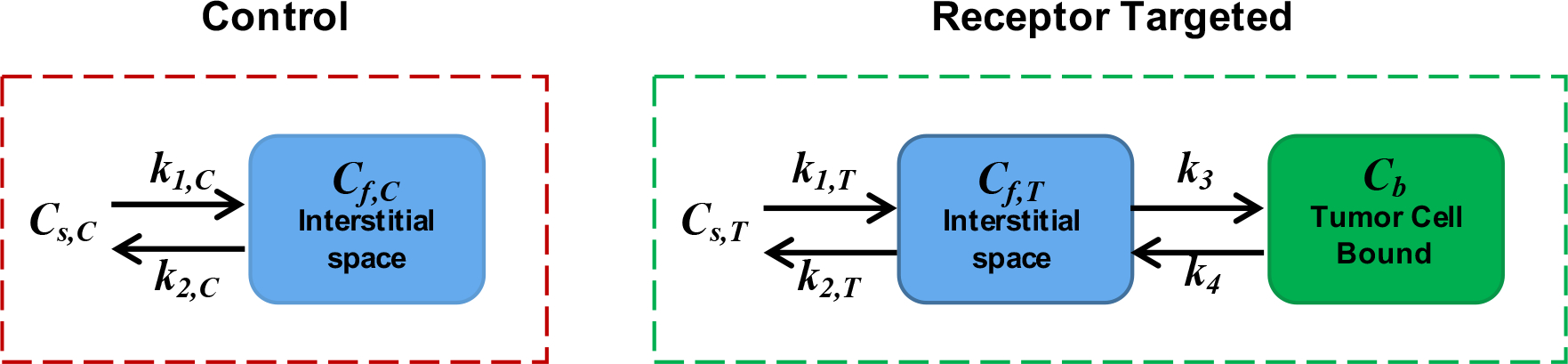
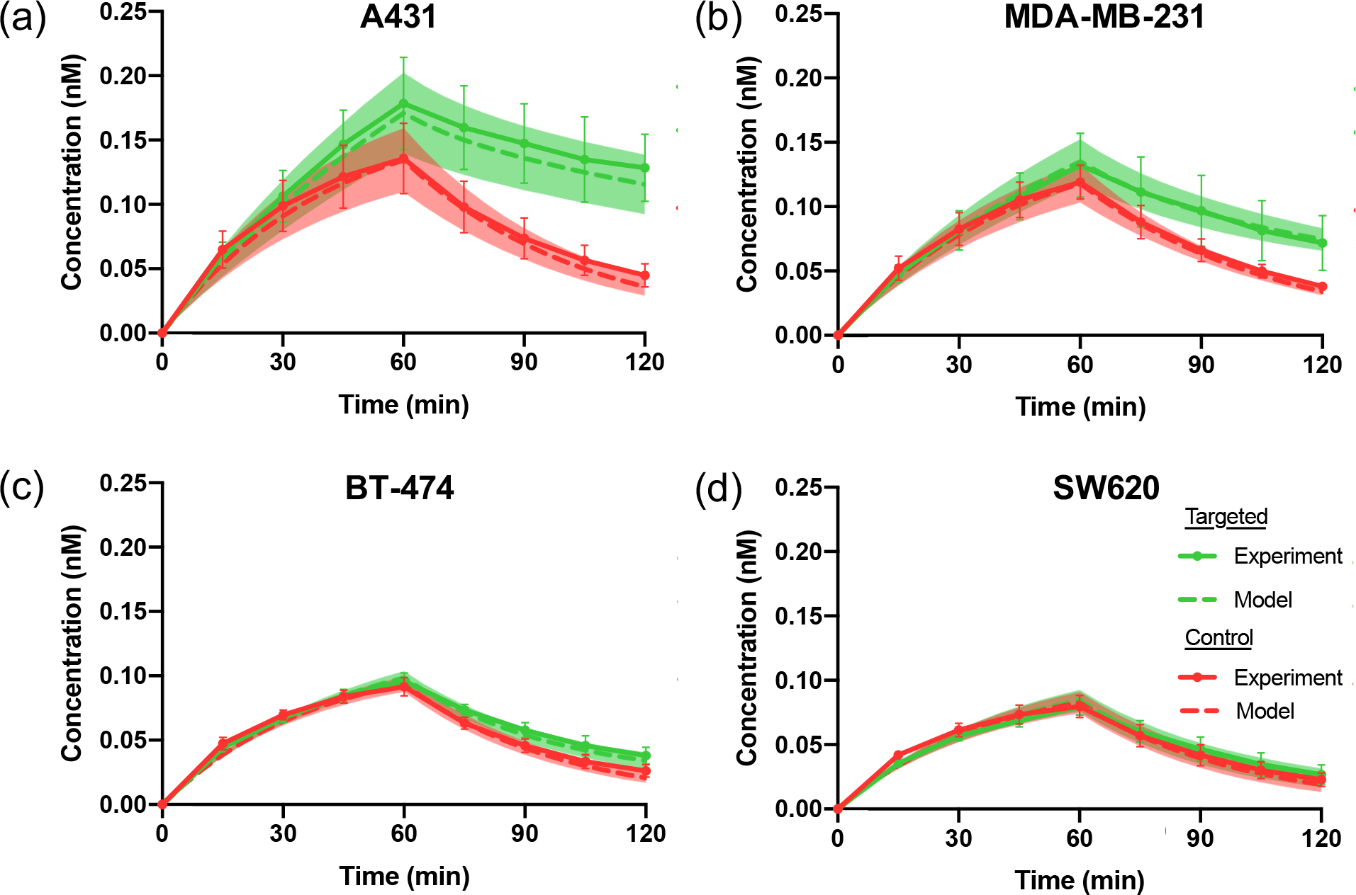
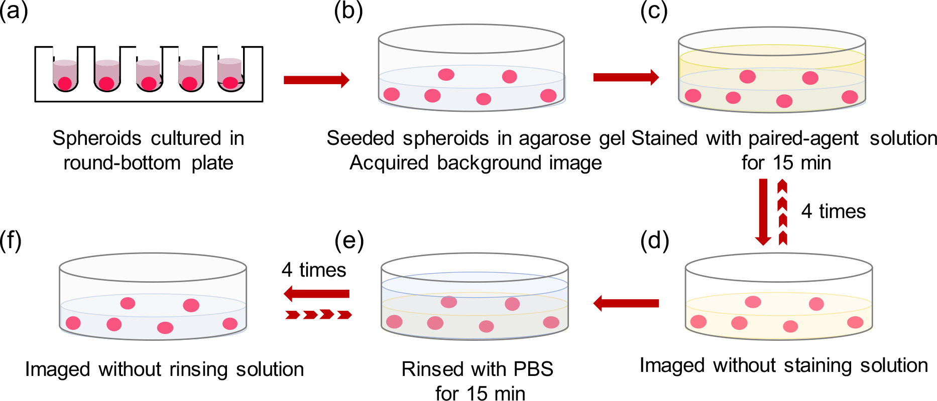
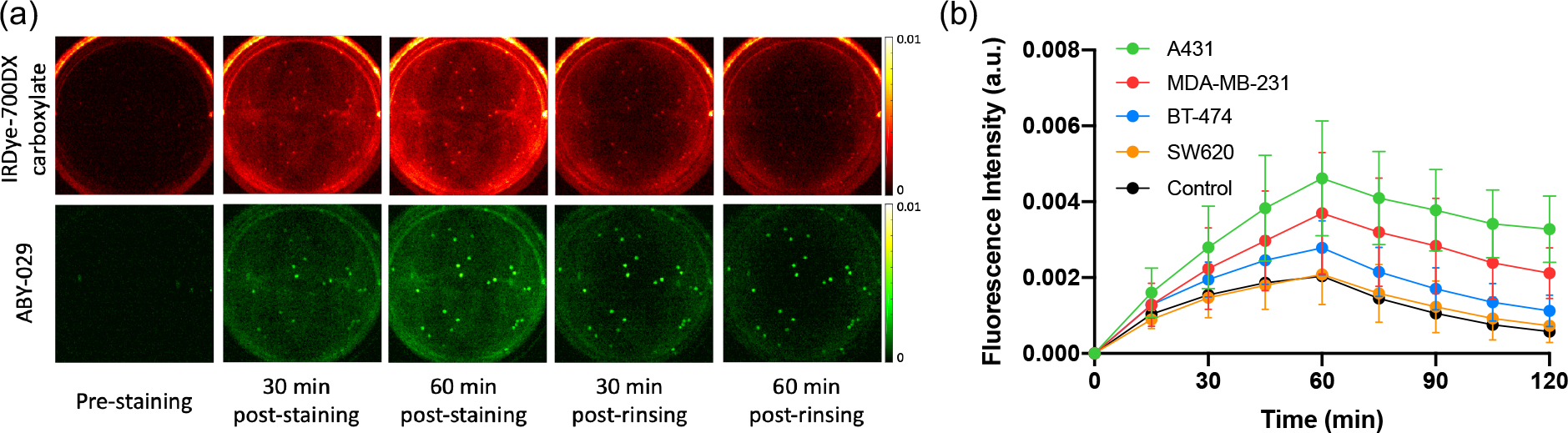
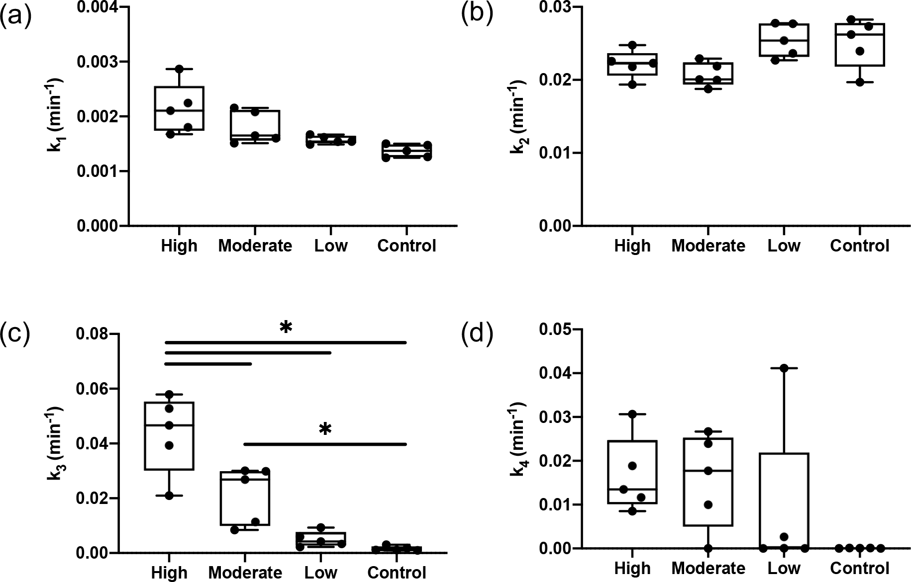
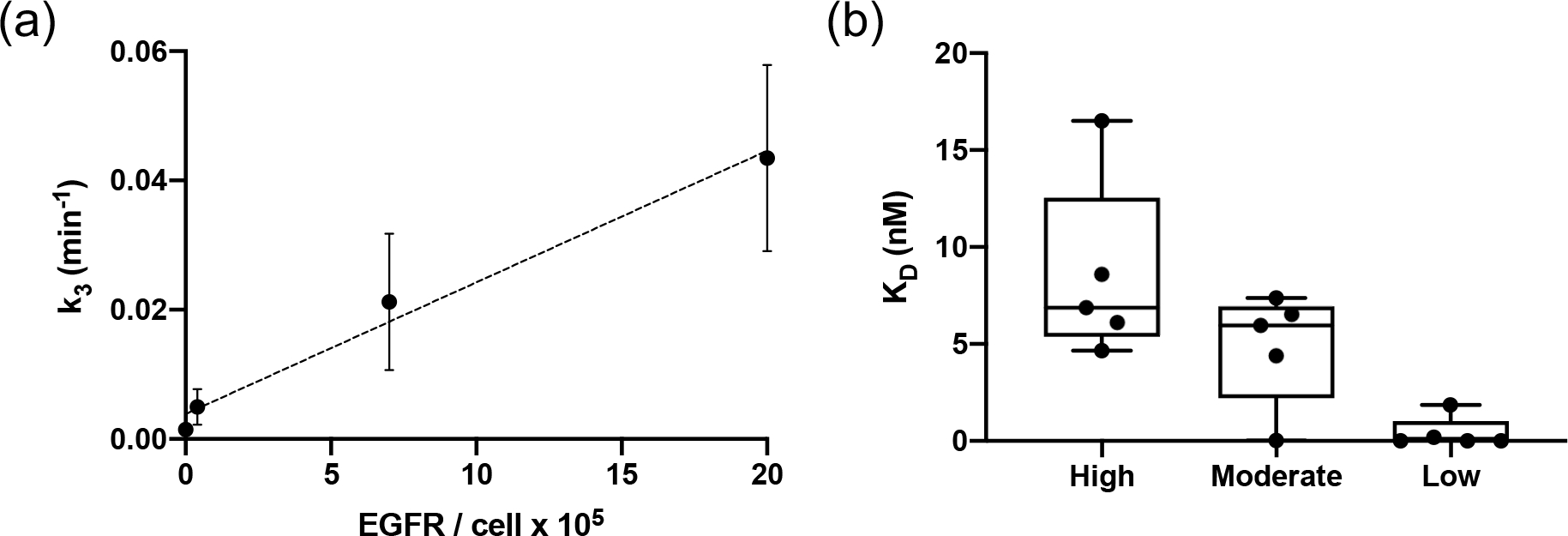
Binding kinetics play an important role in cancer diagnosis and therapeutics. However, current methods of quantifying binding kinetics fail to consider the three-dimensional environment that drugs and imaging agents experience in biological tissue. In response, a methodology to assay agent binding and dissociation in 3-D tissue culture was developed using paired-agent molecular imaging principles. To test the methodology, the uptakes of ABY-029 (an IRDye 800CW-labeled epidermal growth factor receptor (EGFR)-targeted antibody mimetic) and IRDye-700DX carboxylate in 3-D spheroids were measured in four different human cancer cell lines throughout staining and rinsing. A compartment model (optimized for the application) was then fit to the kinetic curves of both imaging agents to estimate binding and dissociation rate constants of the EGFR-targeted ABY-029 agent. A statistically significant correlation was observed between apparent association rate constant (k) and the receptor concentration experimentally and in simulations (r = 0.99, p < 0.05). A statistically significant difference was found between effective k (apparent rate constant of ABY-029 binding to EGFR) values for cell lines with varying levels of EGFR expression (p < 0.05), with no significant difference found between cell lines and controls for other fit parameters. Additionally, a similar binding affinity profile compared to a gold standard method was determined by this model. This low-cost methodology to quantify imaging agent or drug binding affinity in clinically relevant 3-D tumor spheroid models can be used to guide timing of imaging in molecular guided surgery and could have implications in drug development.
结合动力学在癌症诊断和治疗中起着重要作用。然而,目前量化结合动力学的方法未能考虑药物和成像剂在生物组织中所经历的三维环境。因此,开发了一种使用配对剂分子成像原理来测定 3-D 组织培养中结合剂结合和解离的方法。为了测试该方法,在四种不同的人癌细胞系中,通过测量 ABY-029(一种 IRDye 800CW 标记的表皮生长因子受体 (EGFR) 靶向抗体类似物)和 IRDye-700DX 羧酸在 3-D 球体中的摄取量,在整个染色和冲洗过程中对其进行了测试。然后,根据应用情况对动力学曲线进行了优化,以估算 EGFR 靶向 ABY-029 结合剂的结合和解离速率常数。在实验和模拟中,观察到表观结合速率常数 (k) 与受体浓度之间存在显著的相关性 (r = 0.99, p < 0.05)。在具有不同 EGFR 表达水平的细胞系之间,有效 k(ABY-029 与 EGFR 结合的表观速率常数)值存在统计学上的显著差异 (p < 0.05),而对于其他拟合参数,细胞系与对照之间无显著差异。此外,该模型确定了与金标准方法相似的结合亲和力谱。这种在临床相关的 3-D 肿瘤球体模型中定量成像剂或药物结合亲和力的低成本方法可用于指导分子引导手术中的成像时间,并且可能对药物开发产生影响。