HUS Medical Imaging Center, Radiology, University of Helsinki and Helsinki University Hospital.
Folkhälsan Institute of Genetics, Folkhälsan Research Center.
J Hypertens. 2024 Jun 1;42(6):1039-1047. doi: 10.1097/HJH.0000000000003690. Epub 2024 Feb 27.
A third of asymptomatic individuals with type 1 diabetes (T1D) show signs of cerebrovascular disease in brain MRI. These signs associate with advanced stages of diabetic retinal disease, but not in mild or moderate retinopathy. We aimed to evaluate a wider spectrum of retinal changes by exploring the relationship between quantitative measures of retinal vessel parameters (RVP) and cerebrovascular changes in T1D.
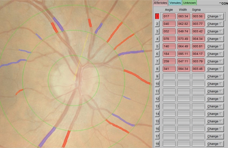
We included 146 neurologically asymptomatic individuals with T1D [51% women, median age 40 (33.0-45.1) years] and 24 healthy, sex-matched and age-matched controls. All individuals underwent a clinical and biochemical work-up and brain MRI, which was evaluated for cerebral microbleeds (CMBs), white matter hyperintensities, and lacunar infarcts. RVPs, including central retinal arteriole (CRAE) and central retinal vein (CRVE) equivalents and the ratio of the two variables (arteriovenous ratio, AVR) were assessed quantitatively by a computer-assisted method (IVAN software, version 3.2.6) from fundus images.
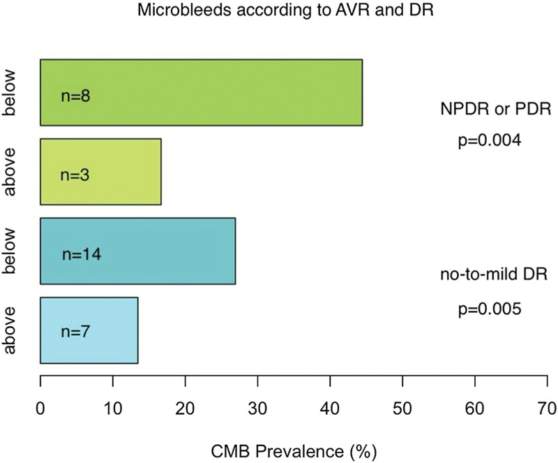
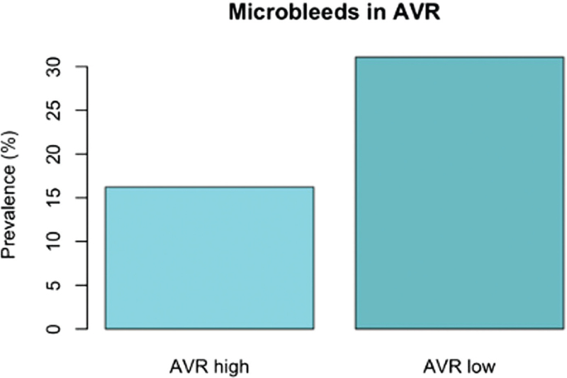
Among T1D participants, those with CMBs had a lower arteriovenous ratio (AVR) compared with those without CMBs ( P = 0.023). AVR was inversely associated with the amount of CMBs ( r = -0.063, P = 0.035). CMB prevalence was higher in those with AVR below the median (31%) compared with above the median (16%, P < 0.001), and this difference was significant also after individuals with only no-to-mild retinopathy were included (28 vs. 16%, P = 0.005). A correlation between blood pressure and CRAE ( r = -0.19, P = 0.025) appeared among those with T1D.
Regardless of the severity of diabetic retinopathy, AVR is associated with the existence of CMBs in T1D.
三分之一的无症状 1 型糖尿病(T1D)患者在脑 MRI 中出现脑血管疾病迹象。这些迹象与糖尿病视网膜病变的晚期阶段有关,但与轻度或中度视网膜病变无关。我们旨在通过探索 T1D 患者视网膜血管参数(RVP)的定量测量值与脑血管变化之间的关系,评估更广泛的视网膜变化谱。
我们纳入了 146 名神经无症状的 T1D 患者(51%为女性,中位年龄 40(33.0-45.1)岁)和 24 名性别和年龄匹配的健康对照者。所有患者均进行了临床和生化检查以及脑部 MRI 检查,以评估脑微出血(CMB)、脑白质高信号和腔隙性梗死。使用计算机辅助方法(IVAN 软件,版本 3.2.6)从眼底图像定量评估 RVPs,包括视网膜中央小动脉(CRAE)和视网膜中央静脉(CRVE)当量以及这两个变量的比值(动静脉比,AVR)。
在 T1D 参与者中,有 CMB 的患者的动静脉比(AVR)低于无 CMB 的患者(P=0.023)。AVR 与 CMB 的数量呈负相关(r=-0.063,P=0.035)。AVR 低于中位数(31%)的患者中 CMB 的患病率高于高于中位数(16%)的患者(P<0.001),而且即使在仅纳入无至轻度视网膜病变的患者后,这种差异仍然显著(28%比 16%,P=0.005)。在 T1D 患者中,血压与 CRAE 之间存在相关性(r=-0.19,P=0.025)。
无论糖尿病视网膜病变的严重程度如何,AVR 与 T1D 患者的 CMB 存在相关。