Programa de Pós-graduação em Microbiologia, Parasitologia e Patologia, Universidade Federal do Paraná, Curitiba, Paraná, Brazil.
EVAHPI Research Group, Laboratório de Biologia Celular, Instituto Carlos Chagas, Fundação Oswaldo Cruz, Curitiba, Paraná, Brazil.
Sci Rep. 2024 Feb 29;14(1):5000. doi: 10.1038/s41598-024-55302-3.
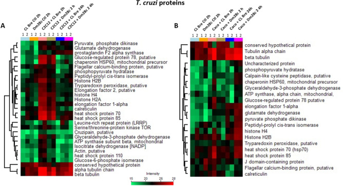
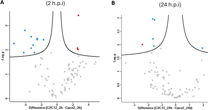
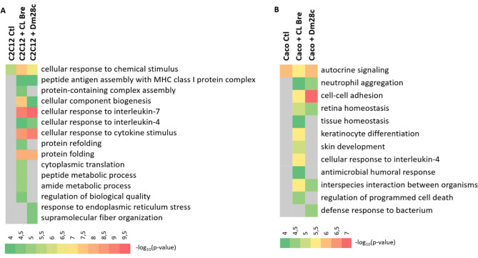
Trypanosoma cruzi is the protozoan that causes Chagas disease (CD), an endemic parasitosis in Latin America distributed around the globe. If CD is not treated in acute phase, the parasite remains silent for years in the host's tissues in a chronic form, which may progress to cardiac, digestive or neurological manifestations. Recently, studies indicated that the gastrointestinal tract represents an important reservoir for T. cruzi in the chronic phase. During interaction T. cruzi and host cells release extracellular vesicles (EVs) that modulates the immune system and infection, but the dynamics of secretion of host and parasite molecules through these EVs is not understood. Now, we used two cell lines: mouse myoblast cell line C2C12, and human intestinal epithelial cell line Caco-2to simulate the environments found by the parasite in the host. We isolated large EVs (LEVs) from the interaction of T. cruzi CL Brener and Dm28c/C2C12 and Caco-2 cells upon 2 and 24 h of infection. Our data showed that at two hours there is a strong cellular response mediated by EVs, both in the number, variety and enrichment/targeting of proteins found in LEVs for diverse functions. Qualitative and quantitative analysis showed that proteins exported in LEVs of C2C12 and Caco-2 have different patterns. We found a predominance of host proteins at early infection. The parasite-host cell interaction induces a switch in the functionality of proteins carried by LEVs and a heterogeneous response depending on the tissues analyzed. Protein-protein interaction analysis showed that cytoplasmic and mitochondrial homologues of the same parasite protein, tryparedoxin peroxidase, were differentially packaged in LEVs, also impacting the interacting molecule of this protein in the host. These data provide new evidence that the interaction with T. cruzi leads to a rapid tissue response through the release of LEVs, reflecting the enrichment of some proteins that could modulate the infection environment.
克氏锥虫是引起恰加斯病(CD)的原生动物,CD 是拉丁美洲流行的地方性寄生虫病,分布于全球各地。如果在急性阶段不治疗 CD,寄生虫会在宿主组织中沉默数年,形成慢性形式,可能会发展为心脏、消化或神经表现。最近的研究表明,胃肠道在慢性期是克氏锥虫的一个重要储存库。在相互作用过程中,克氏锥虫和宿主细胞释放细胞外囊泡(EVs),调节免疫系统和感染,但通过这些 EVs 分泌宿主和寄生虫分子的动力学尚不清楚。现在,我们使用了两种细胞系:小鼠成肌细胞系 C2C12 和人肠道上皮细胞系 Caco-2,来模拟寄生虫在宿主体内发现的环境。我们从 T. cruzi CL Brener 和 Dm28c/C2C12 与 Caco-2 细胞的相互作用中分离出了大细胞外囊泡(LEVs),分别在感染后 2 小时和 24 小时进行。我们的数据表明,在两小时时,EV 介导的细胞反应非常强烈,无论是 LEVs 中发现的蛋白数量、种类,还是其对各种功能的富集/靶向性都很强。定性和定量分析表明,C2C12 和 Caco-2 细胞 LEVs 中输出的蛋白具有不同的模式。我们在早期感染时发现了大量的宿主蛋白。寄生虫-宿主细胞的相互作用会诱导 LEVs 中携带的蛋白的功能发生转变,并根据分析的组织产生异质的反应。蛋白-蛋白相互作用分析表明,同一寄生虫蛋白,即硫氧还蛋白过氧化物酶的细胞质和线粒体同源物,在 LEVs 中被不同地包装,这也影响了该蛋白在宿主中的相互作用分子。这些数据提供了新的证据,表明与 T. cruzi 的相互作用会通过释放 LEVs 导致快速的组织反应,反映了一些可能调节感染环境的蛋白的富集。