• 文献检索
  • 文档翻译
  • 深度研究
  • 学术资讯
  • Suppr Zotero 插件Zotero 插件
  • 邀请有礼
  • 套餐&价格
  • 历史记录
应用&插件
Suppr Zotero 插件Zotero 插件浏览器插件Mac 客户端Windows 客户端微信小程序
定价
高级版会员购买积分包购买API积分包
服务
文献检索文档翻译深度研究API 文档MCP 服务
关于我们
关于 Suppr公司介绍联系我们用户协议隐私条款
关注我们

Suppr 超能文献

核心技术专利:CN118964589B侵权必究
粤ICP备2023148730 号-1Suppr @ 2026

文献检索

告别复杂PubMed语法,用中文像聊天一样搜索,搜遍4000万医学文献。AI智能推荐,让科研检索更轻松。

立即免费搜索

文件翻译

保留排版,准确专业,支持PDF/Word/PPT等文件格式,支持 12+语言互译。

免费翻译文档

深度研究

AI帮你快速写综述,25分钟生成高质量综述,智能提取关键信息,辅助科研写作。

立即免费体验

左束支传导阻滞对心肌 T1 映射的影响。

The influence of left bundle branch block on myocardial T1 mapping.

机构信息

Department of Radiology, Charité-Universitätsmedizin Berlin, corporate member of Freie Universität Berlin and Humboldt-Universität zu Berlin, Hindenburgdamm 30, 12203, Berlin, Germany.

Academic Department of Diagnostic and Interventional Radiology and Paediatric Radiology, Protestant Hospital of the Bethel Foundation, Bielefeld University, Medical School and University Medical Center East Westphalia-Lippe, Burgsteig 13, 33617, Bielefeld, Germany.

出版信息

Sci Rep. 2024 Mar 5;14(1):5379. doi: 10.1038/s41598-024-55821-z.

DOI:10.1038/s41598-024-55821-z
PMID:38438510
原文链接:https://pmc.ncbi.nlm.nih.gov/articles/PMC10912434/
Abstract

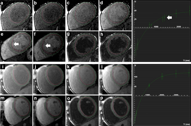
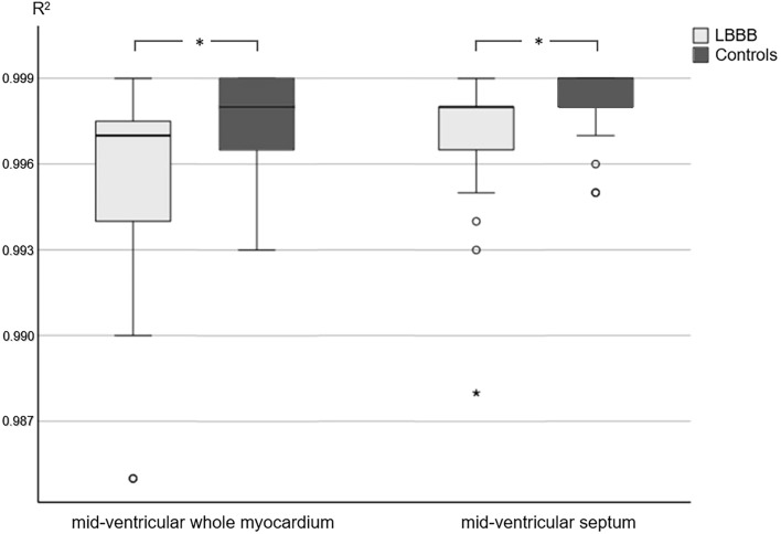
Tissue characterisation using T1 mapping has become an established magnetic resonance imaging (MRI) technique to detect myocardial diseases. This retrospective study aimed to determine the influence of left bundle branch block (LBBB) on T1 mapping at 1.5 T. Datasets of 36 patients with LBBB and 27 healthy controls with T1 mapping (Modified Look-Locker inversion-recovery (MOLLI), 5(3)3 sampling) were included. T1 relaxation times were determined on mid-cavity short-axis images. R maps were generated as a pixel-wise indicator for the goodness of the fit of T1 maps. R values were significantly lower in patients with LBBB than in healthy controls (whole myocardium/septum, 0.997, IQR, 0.00 vs. 0.998, IQR, 0.00; p = 0.008/0.998, IQR, 0.00 vs. 0.999, IQR, 0.00; p = 0.027). Manual correction of semi-automated evaluation tended to improve R values but not significantly. Strain analysis was performed and the systolic dyssynchrony index (SDI) was calculated as a measure for left ventricular dyssynchrony. While MRI is generally prone to artefacts, lower goodness of the fit in LBBB may be mainly attributable to asynchronous contraction. Therefore, careful checking of the source data and, if necessary, manual post-processing is important. New techniques might improve the goodness of the fit of T1 mapping by reducing sampling in the motion prone diastole of LBBB patients.

摘要

使用 T1 映射进行组织特征分析已经成为一种成熟的磁共振成像 (MRI) 技术,可用于检测心肌疾病。本回顾性研究旨在确定左束支传导阻滞 (LBBB) 对 1.5T 下 T1 映射的影响。研究纳入了 36 例 LBBB 患者和 27 例健康对照者的 T1 映射数据集(改良 Look-Locker 反转恢复 (MOLLI),5(3)3 采样)。在心脏中部短轴图像上确定 T1 弛豫时间。R 图作为 T1 图拟合好坏的像素级指示生成。LBBB 患者的 R 值明显低于健康对照组(整个心肌/室间隔,0.997,IQR,0.00 与 0.998,IQR,0.00;p=0.008/0.998,IQR,0.00 与 0.999,IQR,0.00;p=0.027)。半自动评估的手动校正倾向于提高 R 值,但没有统计学意义。进行应变分析,并计算左心室不同步指数 (SDI) 作为左心室不同步的度量。虽然 MRI 通常容易出现伪影,但 LBBB 下拟合度较差可能主要归因于收缩不同步。因此,仔细检查原始数据,如果有必要,进行手动后处理非常重要。新技术可能通过减少 LBBB 患者易受运动影响的舒张期采样来提高 T1 映射的拟合度。

https://cdn.ncbi.nlm.nih.gov/pmc/blobs/bf86/10912434/11dff33b19c0/41598_2024_55821_Fig3_HTML.jpg
https://cdn.ncbi.nlm.nih.gov/pmc/blobs/bf86/10912434/bd23b848385f/41598_2024_55821_Fig1_HTML.jpg
https://cdn.ncbi.nlm.nih.gov/pmc/blobs/bf86/10912434/0424c34b5893/41598_2024_55821_Fig2_HTML.jpg
https://cdn.ncbi.nlm.nih.gov/pmc/blobs/bf86/10912434/11dff33b19c0/41598_2024_55821_Fig3_HTML.jpg
https://cdn.ncbi.nlm.nih.gov/pmc/blobs/bf86/10912434/bd23b848385f/41598_2024_55821_Fig1_HTML.jpg
https://cdn.ncbi.nlm.nih.gov/pmc/blobs/bf86/10912434/0424c34b5893/41598_2024_55821_Fig2_HTML.jpg
https://cdn.ncbi.nlm.nih.gov/pmc/blobs/bf86/10912434/11dff33b19c0/41598_2024_55821_Fig3_HTML.jpg

相似文献

1
The influence of left bundle branch block on myocardial T1 mapping.左束支传导阻滞对心肌 T1 映射的影响。
Sci Rep. 2024 Mar 5;14(1):5379. doi: 10.1038/s41598-024-55821-z.
2
Assessment of left ventricular mechanical dyssynchrony in left bundle branch block canine model: Comparison between cine and tagged MRI.左束支传导阻滞犬模型中左心室机械性不同步的评估:电影磁共振成像与标记磁共振成像的比较
J Magn Reson Imaging. 2016 Oct;44(4):956-63. doi: 10.1002/jmri.25225. Epub 2016 Mar 12.
3
T1 reactivity as an imaging biomarker in myocardial tissue characterization discriminating normal, ischemic and infarcted myocardium.T1反应性作为一种成像生物标志物,用于心肌组织特征描述,以区分正常、缺血和梗死心肌。
Int J Cardiovasc Imaging. 2019 Jul;35(7):1319-1325. doi: 10.1007/s10554-019-01554-4. Epub 2019 May 15.
4
Normal myocardial native T values in children using single-point saturation recovery and modified look-locker inversion recovery (MOLLI).使用单点饱和恢复和改良Look-Locker反转恢复(MOLLI)技术测量儿童正常心肌的固有T值。
J Magn Reson Imaging. 2020 Mar;51(3):897-903. doi: 10.1002/jmri.26910. Epub 2019 Sep 11.
5
Cardiac magnetic resonance imaging in patients with left bundle branch block: Patterns of dyssynchrony and implications for late gadolinium enhancement imaging.左束支传导阻滞患者的心脏磁共振成像:不同步模式及其对延迟钆增强成像的影响。
Front Cardiovasc Med. 2022 Oct 20;9:977414. doi: 10.3389/fcvm.2022.977414. eCollection 2022.
6
Cardiovascular magnetic resonance features of mechanical dyssynchrony in patients with left bundle branch block.左束支传导阻滞患者机械性不同步的心血管磁共振特征
Int J Cardiovasc Imaging. 2016 Sep;32(9):1427-1438. doi: 10.1007/s10554-016-0925-x. Epub 2016 Jun 15.
7
Precision-optimized single protocol pre-/post-contrast modified-look locker inversion T1 mapping using composite inversion group fitting.基于复合反转组拟合的精准优化单协议预/后对比改良 Look-Locker 反转 T1 mapping 技术。
Magn Reson Imaging. 2020 Dec;74:195-202. doi: 10.1016/j.mri.2020.09.025. Epub 2020 Oct 1.
8
Systolic MOLLI T1 mapping with heart-rate-dependent pulse sequence sampling scheme is feasible in patients with atrial fibrillation.采用心率依赖型脉冲序列采样方案的收缩期MOLLI T1映射在心房颤动患者中是可行的。
J Cardiovasc Magn Reson. 2016 Mar 15;18:13. doi: 10.1186/s12968-016-0232-7.
9
The impact of trans-catheter aortic valve replacement induced left-bundle branch block on cardiac reverse remodeling.经导管主动脉瓣置换术所致左束支传导阻滞对心脏逆向重构的影响。
J Cardiovasc Magn Reson. 2017 Feb 22;19(1):22. doi: 10.1186/s12968-017-0335-9.
10
Accelerated cardiac T1 mapping with recurrent networks and cyclic, model-based loss.基于循环、模型的损失和递归网络的加速心脏 T1 映射。
Med Phys. 2022 Nov;49(11):6986-7000. doi: 10.1002/mp.15801. Epub 2022 Jun 21.

引用本文的文献

1
Cardiac Conduction Disorders Due to Acquired or Genetic Causes in Young Adults: A Review of the Current Literature.青年成人后天性或遗传性心脏传导障碍:当前文献综述
J Am Heart Assoc. 2025 May 6;14(9):e040274. doi: 10.1161/JAHA.124.040274. Epub 2025 May 2.

本文引用的文献

1
Comparison of classical Fabry and its p.D313Y and p.A143T variants by cardiac T1 mapping, LGE and feature tracking myocardial strain.采用心脏 T1 映射、LGE 和特征追踪心肌应变技术对经典 Fabry 病及其 p.D313Y 和 p.A143T 变体进行比较。
Sci Rep. 2023 Apr 10;13(1):5809. doi: 10.1038/s41598-023-32464-0.
2
Left ventricular T1-mapping in diastole versus systole in patients with mitral regurgitation.左心室 T1 映射在二尖瓣反流患者的舒张期与收缩期的比较。
Sci Rep. 2022 Nov 21;12(1):20000. doi: 10.1038/s41598-022-23314-6.
3
Cardiac magnetic resonance imaging in patients with left bundle branch block: Patterns of dyssynchrony and implications for late gadolinium enhancement imaging.
左束支传导阻滞患者的心脏磁共振成像:不同步模式及其对延迟钆增强成像的影响。
Front Cardiovasc Med. 2022 Oct 20;9:977414. doi: 10.3389/fcvm.2022.977414. eCollection 2022.
4
T1 values and extracellular volume fraction in asymptomatic subjects: variations in left ventricular segments and correlation with cardiovascular risk factors.无症状受试者的 T1 值和细胞外容积分数:左心室节段的变化与心血管危险因素的相关性。
Sci Rep. 2022 Jul 22;12(1):12544. doi: 10.1038/s41598-022-16696-0.
5
Accelerated cardiac T1 mapping with recurrent networks and cyclic, model-based loss.基于循环、模型的损失和递归网络的加速心脏 T1 映射。
Med Phys. 2022 Nov;49(11):6986-7000. doi: 10.1002/mp.15801. Epub 2022 Jun 21.
6
Using multi-parametric quantitative MRI to screen for cardiac involvement in patients with idiopathic inflammatory myopathy.采用多参数定量 MRI 筛查特发性炎性肌病患者的心脏受累情况。
Sci Rep. 2022 Jun 14;12(1):9819. doi: 10.1038/s41598-022-13858-y.
7
The Road Toward Reproducibility of Parametric Mapping of the Heart: A Technical Review.心脏参数映射的可重复性之路:技术综述
Front Cardiovasc Med. 2022 May 6;9:876475. doi: 10.3389/fcvm.2022.876475. eCollection 2022.
8
Myocardial T1 Values at 1.5 T: Normal Values for General Electric Scanners and Sex-Related Differences.1.5T 下心肌 T1 值:通用电气扫描仪的正常值及性别差异。
J Magn Reson Imaging. 2021 Nov;54(5):1486-1500. doi: 10.1002/jmri.27639. Epub 2021 Apr 13.
9
Morphologies and prognostic significance of left ventricular volume/time curves with cardiac magnetic resonance in patients with non-ischaemic heart failure and left bundle branch block.左心室容积/时间曲线形态及其在非缺血性心力衰竭伴左束支传导阻滞患者心脏磁共振中的预后意义。
Int J Cardiovasc Imaging. 2021 Jul;37(7):2245-2255. doi: 10.1007/s10554-021-02194-3. Epub 2021 Feb 26.
10
Deep learning with attention supervision for automated motion artefact detection in quality control of cardiac T1-mapping.用于心脏T1映射质量控制中自动运动伪影检测的带注意力监督的深度学习
Artif Intell Med. 2020 Nov;110:101955. doi: 10.1016/j.artmed.2020.101955. Epub 2020 Sep 7.