• 文献检索
  • 文档翻译
  • 深度研究
  • 学术资讯
  • Suppr Zotero 插件Zotero 插件
  • 邀请有礼
  • 套餐&价格
  • 历史记录
应用&插件
Suppr Zotero 插件Zotero 插件浏览器插件Mac 客户端Windows 客户端微信小程序
定价
高级版会员购买积分包购买API积分包
服务
文献检索文档翻译深度研究API 文档MCP 服务
关于我们
关于 Suppr公司介绍联系我们用户协议隐私条款
关注我们

Suppr 超能文献

核心技术专利:CN118964589B侵权必究
粤ICP备2023148730 号-1Suppr @ 2026

文献检索

告别复杂PubMed语法,用中文像聊天一样搜索,搜遍4000万医学文献。AI智能推荐,让科研检索更轻松。

立即免费搜索

文件翻译

保留排版,准确专业,支持PDF/Word/PPT等文件格式,支持 12+语言互译。

免费翻译文档

深度研究

AI帮你快速写综述,25分钟生成高质量综述,智能提取关键信息,辅助科研写作。

立即免费体验

原发性侧索硬化症患者的脑-小脑连接性增强。

Cerebro-cerebellar connectivity is increased in primary lateral sclerosis.

作者信息

Meoded Avner, Morrissette Arthur E, Katipally Rohan, Schanz Olivia, Gotts Stephen J, Floeter Mary Kay

机构信息

National Institute of Neurological Disorders and Stroke, National Institutes of Health, Bethesda, MD, USA.

National Institute of Mental Health, National Institutes of Health, Bethesda, MD, USA.

出版信息

Neuroimage Clin. 2014 Dec 9;7:288-96. doi: 10.1016/j.nicl.2014.12.009. eCollection 2015.

DOI:10.1016/j.nicl.2014.12.009
PMID:25610792
原文链接:https://pmc.ncbi.nlm.nih.gov/articles/PMC4300015/
Abstract

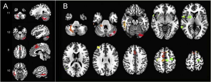
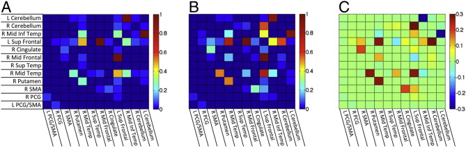
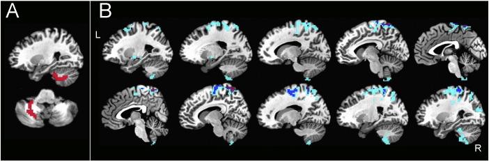
Increased functional connectivity in resting state networks was found in several studies of patients with motor neuron disorders, although diffusion tensor imaging studies consistently show loss of white matter integrity. To understand the relationship between structural connectivity and functional connectivity, we examined the structural connections between regions with altered functional connectivity in patients with primary lateral sclerosis (PLS), a long-lived motor neuron disease. Connectivity matrices were constructed from resting state fMRI in 16 PLS patients to identify areas of differing connectivity between patients and healthy controls. Probabilistic fiber tracking was used to examine structural connections between regions of differing connectivity. PLS patients had 12 regions with increased functional connectivity compared to controls, with a predominance of cerebro-cerebellar connections. Increased functional connectivity was strongest between the cerebellum and cortical motor areas and between the cerebellum and frontal and temporal cortex. Fiber tracking detected no difference in connections between regions with increased functional connectivity. We conclude that functional connectivity changes are not strongly based in structural connectivity. Increased functional connectivity may be caused by common inputs, or by reduced selectivity of cortical activation, which could result from loss of intracortical inhibition when cortical afferents are intact.

摘要

在多项针对运动神经元疾病患者的研究中,发现静息态网络中的功能连接性增强,尽管扩散张量成像研究一直显示白质完整性受损。为了理解结构连接性与功能连接性之间的关系,我们研究了原发性侧索硬化症(PLS,一种病程较长的运动神经元疾病)患者中功能连接性发生改变的区域之间的结构连接。从16例PLS患者的静息态功能磁共振成像构建连接矩阵,以识别患者与健康对照之间连接性不同的区域。使用概率纤维追踪来检查连接性不同的区域之间的结构连接。与对照组相比,PLS患者有12个区域的功能连接性增强,以脑-小脑连接为主。小脑与皮质运动区之间以及小脑与额叶和颞叶皮质之间的功能连接性增强最为明显。纤维追踪未发现功能连接性增强区域之间的连接存在差异。我们得出结论,功能连接性变化并非强烈基于结构连接性。功能连接性增加可能是由共同输入引起的,或者是由皮质激活的选择性降低引起的,当皮质传入完整时,这可能是由于皮质内抑制丧失所致。

https://cdn.ncbi.nlm.nih.gov/pmc/blobs/2d20/4300015/60e7363d3321/gr6.jpg
https://cdn.ncbi.nlm.nih.gov/pmc/blobs/2d20/4300015/da3e5ca0f296/gr1.jpg
https://cdn.ncbi.nlm.nih.gov/pmc/blobs/2d20/4300015/7012dceaa0f8/gr2.jpg
https://cdn.ncbi.nlm.nih.gov/pmc/blobs/2d20/4300015/8b0e08b5cf95/gr4.jpg
https://cdn.ncbi.nlm.nih.gov/pmc/blobs/2d20/4300015/2e23bb306ece/gr5.jpg
https://cdn.ncbi.nlm.nih.gov/pmc/blobs/2d20/4300015/60e7363d3321/gr6.jpg

相似文献

1
Cerebro-cerebellar connectivity is increased in primary lateral sclerosis.原发性侧索硬化症患者的脑-小脑连接性增强。
Neuroimage Clin. 2014 Dec 9;7:288-96. doi: 10.1016/j.nicl.2014.12.009. eCollection 2015.
2
Fibromyalgia is characterized by altered frontal and cerebellar structural covariance brain networks.纤维肌痛的特征是额叶和小脑结构协方差脑网络改变。
Neuroimage Clin. 2015 Mar 4;7:667-77. doi: 10.1016/j.nicl.2015.02.022. eCollection 2015.
3
Interhemispheric connectivity in amyotrophic lateral sclerosis: A near-infrared spectroscopy and diffusion tensor imaging study.肌萎缩侧索硬化症的半球间连接性:一项近红外光谱和扩散张量成像研究。
Neuroimage Clin. 2016 Sep 29;12:666-672. doi: 10.1016/j.nicl.2016.09.020. eCollection 2016.
4
Structural imaging differences and longitudinal changes in primary lateral sclerosis and amyotrophic lateral sclerosis.原发性侧索硬化症和肌萎缩性侧索硬化症的结构影像学差异和纵向变化。
Neuroimage Clin. 2012 Dec 24;2:151-60. doi: 10.1016/j.nicl.2012.12.003. eCollection 2012.
5
The two-year progression of structural and functional cerebral MRI in amyotrophic lateral sclerosis.肌萎缩侧索硬化症的结构和功能大脑 MRI 的两年进展。
Neuroimage Clin. 2017 Dec 18;17:953-961. doi: 10.1016/j.nicl.2017.12.025. eCollection 2018.
6
Supra- and infra-tentorial degeneration patterns in primary lateral sclerosis: a multimodal longitudinal neuroradiology study.原发性侧索硬化症的幕上和幕下退行性变模式:一项多模态纵向神经影像学研究。
J Neurol. 2024 Jun;271(6):3239-3255. doi: 10.1007/s00415-024-12261-z. Epub 2024 Mar 5.
7
Loss of functional connectivity is an early imaging marker in primary lateral sclerosis.功能连接性丧失是原发性侧索硬化症的一种早期影像学标志物。
Amyotroph Lateral Scler Frontotemporal Degener. 2018 Nov;19(7-8):562-569. doi: 10.1080/21678421.2018.1517180. Epub 2018 Oct 9.
8
Involvement of the dentate nucleus in the pathophysiology of amyotrophic lateral sclerosis: A multi-center and multi-modal neuroimaging study.齿状核在肌萎缩侧索硬化症病理生理学中的作用:一项多中心多模态神经影像学研究。
Neuroimage Clin. 2020;28:102385. doi: 10.1016/j.nicl.2020.102385. Epub 2020 Aug 16.
9
Reduced structural connectivity within a prefrontal-motor-subcortical network in amyotrophic lateral sclerosis.肌萎缩侧索硬化症患者前额叶-运动-皮质下网络内的结构连接性降低。
J Magn Reson Imaging. 2015 May;41(5):1342-52. doi: 10.1002/jmri.24695. Epub 2014 Jul 14.
10
Differential involvement of corticospinal tract (CST) fibers in UMN-predominant ALS patients with or without CST hyperintensity: A diffusion tensor tractography study.皮质脊髓束(CST)纤维在伴有或不伴有CST高信号的以UMN为主的肌萎缩侧索硬化症患者中的差异参与:一项扩散张量纤维束成像研究。
Neuroimage Clin. 2017 Feb 22;14:574-579. doi: 10.1016/j.nicl.2017.02.017. eCollection 2017.

引用本文的文献

1
Brain White Matter Changes in Non-demented Individuals with Color Discrimination Deficits and Their Association with Cognitive Impairment: A NODDI Study.色觉辨别缺陷的非痴呆个体脑白质变化及其与认知障碍的关联:一项基于神经突方向离散度与密度成像(NODDI)的研究
Neurosci Bull. 2025 Mar 7. doi: 10.1007/s12264-025-01373-9.
2
Cerebellum in Alzheimer's disease and other neurodegenerative diseases: an emerging research frontier.阿尔茨海默病及其他神经退行性疾病中的小脑:一个新兴的研究前沿。
MedComm (2020). 2024 Jul 13;5(7):e638. doi: 10.1002/mco2.638. eCollection 2024 Jul.
3
Supra- and infra-tentorial degeneration patterns in primary lateral sclerosis: a multimodal longitudinal neuroradiology study.

本文引用的文献

1
Correlation between structural and functional connectivity impairment in amyotrophic lateral sclerosis.肌萎缩侧索硬化症中结构和功能连接损害之间的相关性。
Hum Brain Mapp. 2014 Sep;35(9):4386-95. doi: 10.1002/hbm.22481. Epub 2014 Mar 6.
2
Effective Preprocessing Procedures Virtually Eliminate Distance-Dependent Motion Artifacts in Resting State FMRI.有效的预处理程序几乎可以消除静息态功能磁共振成像中与距离相关的运动伪影。
J Appl Math. 2013 May 21;2013. doi: 10.1155/2013/935154.
3
Alterations in regional functional coherence within the sensory-motor network in amyotrophic lateral sclerosis.
原发性侧索硬化症的幕上和幕下退行性变模式:一项多模态纵向神经影像学研究。
J Neurol. 2024 Jun;271(6):3239-3255. doi: 10.1007/s00415-024-12261-z. Epub 2024 Mar 5.
4
Primary Lateral Sclerosis: An Overview.原发性侧索硬化症概述
J Clin Med. 2024 Jan 19;13(2):578. doi: 10.3390/jcm13020578.
5
Cortico-muscular coherence in primary lateral sclerosis reveals abnormal cortical engagement during motor function beyond primary motor areas.原发性侧索硬化症中的皮质-肌肉相干性揭示了运动功能中初级运动区以外的皮质异常参与。
Cereb Cortex. 2023 Jun 20;33(13):8712-8723. doi: 10.1093/cercor/bhad152.
6
Cerebellar pathology in motor neuron disease: neuroplasticity and neurodegeneration.运动神经元病中的小脑病理学:神经可塑性与神经变性
Neural Regen Res. 2022 Nov;17(11):2335-2341. doi: 10.4103/1673-5374.336139.
7
Brain Connectivity and Network Analysis in Amyotrophic Lateral Sclerosis.肌萎缩侧索硬化症中的脑连接性与网络分析
Neurol Res Int. 2022 Feb 7;2022:1838682. doi: 10.1155/2022/1838682. eCollection 2022.
8
Classification of amyotrophic lateral sclerosis by brain volume, connectivity, and network dynamics.基于脑容量、连接和网络动力学的肌萎缩侧索硬化分类。
Hum Brain Mapp. 2022 Feb 1;43(2):681-699. doi: 10.1002/hbm.25679. Epub 2021 Oct 16.
9
Frontotemporal Pathology in Motor Neuron Disease Phenotypes: Insights From Neuroimaging.运动神经元病表型中的额颞叶病理学:神经影像学见解
Front Neurol. 2021 Aug 16;12:723450. doi: 10.3389/fneur.2021.723450. eCollection 2021.
10
Enhanced inter-regional coupling of neural responses and repetition suppression provide separate contributions to long-term behavioral priming.增强的神经反应区域间耦合和重复抑制为长期行为启动提供了独立的贡献。
Commun Biol. 2021 Apr 20;4(1):487. doi: 10.1038/s42003-021-02002-7.
肌萎缩侧索硬化症患者感觉运动网络内区域功能连贯性的改变。
Neurosci Lett. 2014 Jan 13;558:192-6. doi: 10.1016/j.neulet.2013.11.022. Epub 2013 Nov 22.
4
Amyotrophic lateral sclerosis--a model of corticofugal axonal spread.肌萎缩性侧索硬化症——皮质传出轴突扩散的模型。
Nat Rev Neurol. 2013 Dec;9(12):708-14. doi: 10.1038/nrneurol.2013.221. Epub 2013 Nov 12.
5
Functional connectivity and graph theory in preclinical Alzheimer's disease.临床前阿尔茨海默病中的功能连接性与图论
Neurobiol Aging. 2014 Apr;35(4):757-68. doi: 10.1016/j.neurobiolaging.2013.10.081. Epub 2013 Oct 18.
6
Resting state functional connectivity alterations in primary lateral sclerosis.原发性侧索硬化症静息态功能连接的改变
Neurobiol Aging. 2014 Apr;35(4):916-25. doi: 10.1016/j.neurobiolaging.2013.09.041. Epub 2013 Oct 12.
7
Functional connectivity changes differ in early and late-onset Alzheimer's disease.早期和晚期发病的阿尔茨海默病的功能连接变化不同。
Hum Brain Mapp. 2014 Jul;35(7):2978-94. doi: 10.1002/hbm.22379. Epub 2013 Oct 5.
8
Two distinct forms of functional lateralization in the human brain.人类大脑中两种截然不同的功能偏侧化形式。
Proc Natl Acad Sci U S A. 2013 Sep 3;110(36):E3435-44. doi: 10.1073/pnas.1302581110. Epub 2013 Aug 19.
9
The involvement of the cerebellum in amyotrophic lateral sclerosis.小脑在肌萎缩侧索硬化症中的作用。
Amyotroph Lateral Scler Frontotemporal Degener. 2013 Dec;14(7-8):507-15. doi: 10.3109/21678421.2013.812661. Epub 2013 Jul 29.
10
Correcting brain-wide correlation differences in resting-state FMRI.校正静息态 fMRI 中的全脑相关差异。
Brain Connect. 2013;3(4):339-52. doi: 10.1089/brain.2013.0156. Epub 2013 Jul 31.