Cho Hyun-Kyung, Kee Changwon
Department of Ophthalmology, Gyeongsang National University Changwon Hospital, Gyeongsang National University, School of Medicine, Changwon, Republic of Korea.
Institute of Health Sciences, School of Medicine, Gyeongsang National University, Jinju, Republic of Korea.
J Ophthalmol. 2024 Feb 27;2024:9978354. doi: 10.1155/2024/9978354. eCollection 2024.
This study aimed to investigate longitudinal rates of change (LRCs) of structural parameters from optical coherence tomography (OCT) in patients with primary angle closure glaucoma (PACG) after laser iridotomy (LI) along with laser peripheral iridoplasty (PI).
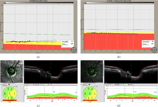
Among 146 patients diagnosed with PACG, thirty-two subjects (32 eyes) who underwent LI plus PI and accomplished more than five times of reliable OCT tests were included in the current retrospective study. Retinal nerve fiber layer (RNFL) and Bruch's membrane opening-minimum rim width (BMO-MRW) were measured by spectral-domain OCT with three month interval. LRCs of global and six Garway-Heath sectors were investigated using the linear mixed-effects model which adjusted BMO area, sex, and age. Imaging of dual Scheimpflug analyzer was performed before and at 1 week after LI with PI and yearly thereafter.
The mean follow-up period was 32.28 ± 13.34 months with a mean number of 10.18 ± 3.33 OCT images. Baseline characteristics are as follows: age, 63 ± 7.9 years; female, 62.5%; intraocular pressure(IOP), 15.48 ± 4.79 mmHg; anterior chamber depth, 2.09 ± 0.18 mm; and mean deviation, -7.97 ± 8.48 dB. Global LRC of BMO-MRW was 0.86 ± 1.34 m/yr and RNFL was -0.64 ± 0.22 m/yr. IOP decreased significantly to 13.06 ± 2.21 mmHg (=0.001) while anterior chamber volume (=0.011) and mean anterior chamber angle (=0.022) increased significantly after LI along with PI compared to the baseline at the final visit.
LRC of a new parameter, BMO-MRW, and LRC of RNFL were relatively low in patients with PACG, following LI along with PI. After widening of the anterior chamber angle and decrease of IOP due to LI plus PI, PACG might show stable structural prognosis assessed by OCT.
本研究旨在调查原发性闭角型青光眼(PACG)患者在接受激光虹膜切开术(LI)联合激光周边虹膜成形术(PI)后,光学相干断层扫描(OCT)测量的结构参数的纵向变化率(LRC)。
在146例诊断为PACG的患者中,本回顾性研究纳入了32例接受LI加PI且完成了超过5次可靠OCT检查的受试者(32只眼)。采用光谱域OCT每隔3个月测量视网膜神经纤维层(RNFL)和布鲁赫膜开口最小边缘宽度(BMO-MRW)。使用调整了BMO面积、性别和年龄的线性混合效应模型研究整体及六个Garway-Heath扇形区域的LRC。在LI联合PI术前、术后1周及此后每年进行双Scheimpflug分析仪成像。
平均随访期为32.28±13.34个月,平均OCT图像数量为10.18±3.33张。基线特征如下:年龄63±7.9岁;女性占62.5%;眼压(IOP)15.48±4.79 mmHg;前房深度2.09±0.18 mm;平均偏差-7.97±8.48 dB。BMO-MRW的整体LRC为0.86±1.34μm/年,RNFL为-0.64±0.22μm/年。与末次随访时的基线相比,LI联合PI术后眼压显著降至13.06±2.21 mmHg(P=0.001),前房容积(P=0.011)和平均前房角(P=0.022)显著增加。
在接受LI联合PI的PACG患者中,新参数BMO-MRW的LRC和RNFL的LRC相对较低。LI联合PI使前房角增宽、眼压降低后,PACG通过OCT评估可能显示出稳定的结构预后。