Tanaka Hiroki, Koyasu Sho, Kikuchi Masahiro, Iima Mami, Omori Koichi, Nakamoto Yuji
Department of Diagnostic Imaging and Nuclear Medicine, Graduate School of Medicine, Kyoto University, Kyoto, Kyoto, Japan.
Department of Otolaryngology, Head and Neck Surgery, Graduate School of Medicine, Kyoto University, Kyoto, Kyoto, Japan.
Magn Reson Med Sci. 2025 Apr 1;24(2):210-219. doi: 10.2463/mrms.mp.2023-0137. Epub 2024 Mar 7.
The 8th edition of the American Joint Committee on Cancer staging system included the depth of invasion (DOI) for the T classification of oral cancer. However, no standardized method has been established to clinically measure the DOI. This study aimed to investigate the accuracy of MRI-based DOI for oral tongue squamous cell carcinoma (OTSCC) in each MRI sequence.
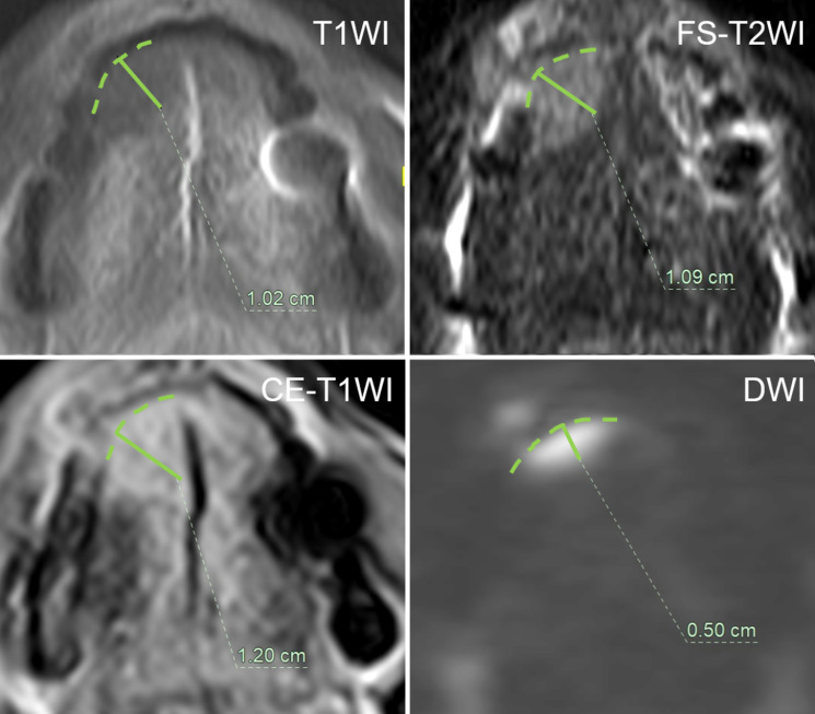
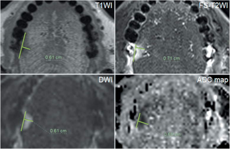
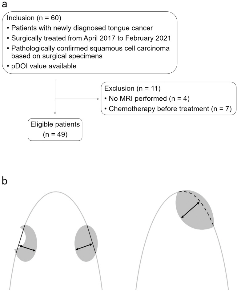
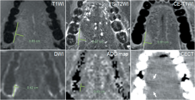
We enrolled 49 patients with histologically proven OTSCC, treated surgically between April 2017 and February 2021. We divided the DOI into three groups using 5 and 10 mm, the thresholds for determining the T stage, and retrospectively evaluated the agreement between MRI-based DOI and pathological DOI (pDOI) for each MRI sequence, axial T1-weighted imaging (T1WI), T2-weighted imaging with fat suppression (FS-T2WI), contrast-enhanced T1WI with fat suppression (CE-T1WI), diffusion-weighted imaging (DWI), and apparent diffusion coefficient (ADC) maps. We also divided the DOI into two groups using 3 mm, the threshold for considering elective neck dissection, and evaluated the overestimation rate of MRI-based DOI in lesions with pDOI ≤ 3 mm.
With 5-mm and 10-mm divisions, the accuracy of the DOI assessment was highest on DWI (0.82, weighted kappa = 0.85). With a 3-mm division, the accuracy was also highest on DWI (0.87, kappa = 0.73). The overestimation rate of the MRI-based DOI in lesions with pDOI ≤ 3 mm was lowest on DWI (27.8%).
DOI on DWI exhibits a comparatively higher rate of concordance with pDOI. DWI may be more useful than other MRI sequences in evaluating the DOI of OTSCC.
美国癌症联合委员会第8版分期系统纳入了口腔癌T分类的浸润深度(DOI)。然而,尚未建立临床测量DOI的标准化方法。本研究旨在探讨基于MRI的DOI在各MRI序列中对口腔舌鳞状细胞癌(OTSCC)的准确性。
我们纳入了49例经组织学证实的OTSCC患者,这些患者在2017年4月至2021年2月期间接受了手术治疗。我们使用5毫米和10毫米(确定T分期的阈值)将DOI分为三组,并回顾性评估了基于MRI的DOI与各MRI序列(轴位T1加权成像(T1WI)、脂肪抑制T2加权成像(FS-T2WI)、脂肪抑制对比增强T1WI(CE-T1WI)、扩散加权成像(DWI)和表观扩散系数(ADC)图)的病理DOI(pDOI)之间的一致性。我们还使用3毫米(考虑选择性颈清扫的阈值)将DOI分为两组,并评估了pDOI≤3毫米的病变中基于MRI的DOI的高估率。
以5毫米和10毫米划分时,DOI评估的准确性在DWI上最高(0.82,加权kappa=0.85)。以3毫米划分时,准确性在DWI上也最高(0.87,kappa=0.73)。在pDOI≤3毫米的病变中,基于MRI的DOI的高估率在DWI上最低(27.8%)。
DWI上的DOI与pDOI表现出相对较高的一致性。在评估OTSCC的DOI方面,DWI可能比其他MRI序列更有用。