Department of Pathology, Tianjin Union Medical Center, Tianjin 300121, P.R. China.
Graduate School, Tianjin Medical University, Tianjin 300070, P.R. China.
Oncol Rep. 2024 May;51(5). doi: 10.3892/or.2024.8722. Epub 2024 Mar 8.
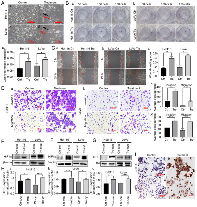
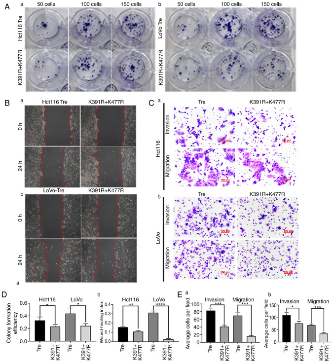
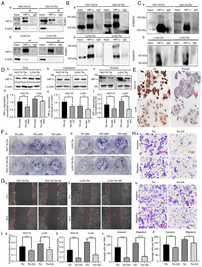
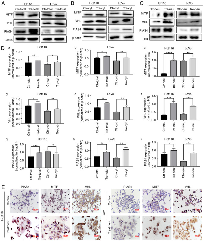
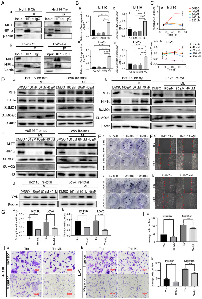
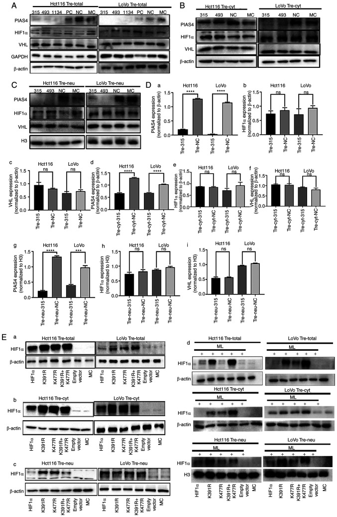
High concentrations of cobalt chloride (CoCl) can induce the formation of polyploid giant cancer cells (PGCCs) in various tumors, which can produce daughter cells with strong proliferative, migratory and invasive abilities via asymmetric division. To study the role of hypoxia‑inducible factor (HIF) 1α in the formation of PGCCs, colon cancer cell lines Hct116 and LoVo were used as experimental subjects. Western blotting, nuclear and cytoplasmic protein extraction and immunocytochemical experiments were used to compare the changes in the expression and subcellular localization of HIF1α, microphthalmia‑associated transcription factor (MITF), protein inhibitor of activated STAT protein 4 (PIAS4) and von Hippel‑Lindau disease tumor suppressor (VHL) after treatment with CoCl. The SUMOylation of HIFα was verified by co‑immunoprecipitation assay. After inhibiting HIF1α SUMOylation, the changes in proliferation, migration and invasion abilities of Hct116 and LoVo were compared by plate colony formation, wound healing and Transwell migration and invasion. In addition, lysine sites that led to SUMOylation of HIF1α were identified through site mutation experiments. The results showed that CoCl can induce the formation of PGCCs with the expression level of HIF1α higher in treated cells than in control cells. HIF1α was primarily located in the cytoplasm of control cell. Following CoCl treatment, the subcellular localization of HIF1α was primarily in the nuclei of PGCCs with daughter cells (PDCs). After treatment with SUMOylation inhibitors, the nuclear HIF1α expression in PDCs decreased. Furthermore, their proliferation, migration and invasion abilities also decreased. After inhibiting the expression of MITF, the expression of HIF1α decreased. MITF can regulate HIF1α SUMOylation. Expression and subcellular localization of VHL and HIF1α did not change following PIAS4 knockdown. SUMOylation of HIF1α occurs at the amino acid sites K391 and K477 in PDCs. After mutation of the two sites, nuclear expression of HIF1α in PDCs was reduced, along with a significant reduction in the proliferation, migration and invasion abilities. In conclusion, the post‑translation modification regulated the subcellular location of HIF1α and the nuclear expression of HIF1α promoted the proliferation, migration and invasion abilities of PDCs. MITF could regulate the transcription and protein levels of HIF1α and participate in the regulation of HIF1α SUMOylation.
高浓度的氯化钴(CoCl)可诱导多种肿瘤中多倍体巨大癌细胞(PGCC)的形成,这些细胞可通过不对称分裂产生具有较强增殖、迁移和侵袭能力的子细胞。为了研究缺氧诱导因子(HIF)1α在 PGCC 形成中的作用,以结肠癌细胞系 Hct116 和 LoVo 作为实验对象。采用 Western blot 法、核质蛋白提取和免疫细胞化学实验,比较 CoCl 处理后 HIF1α、小眼畸形相关转录因子(MITF)、信号转导和转录激活因子 4 蛋白抑制剂(PIAS4)和 von Hippel-Lindau 疾病肿瘤抑制因子(VHL)的表达和亚细胞定位变化。通过共免疫沉淀实验验证 HIFα的 SUMO 化。抑制 HIF1α SUMO 化后,通过平板集落形成、划痕愈合和 Transwell 迁移及侵袭实验比较 Hct116 和 LoVo 增殖、迁移和侵袭能力的变化。此外,通过位点突变实验鉴定导致 HIF1α SUMO 化的赖氨酸位点。结果显示,CoCl 可诱导 PGCC 形成,与对照组相比,处理组细胞 HIF1α 的表达水平更高。对照组细胞中 HIF1α 主要位于细胞质中。CoCl 处理后,HIF1α 的亚细胞定位主要在具有子细胞(PDCs)的 PGCC 核内。SUMO 化抑制剂处理后,PDC 核内 HIF1α 表达减少,其增殖、迁移和侵袭能力也随之下降。抑制 MITF 表达后,HIF1α 表达减少。MITF 可调节 HIF1α SUMO 化。敲低 PIAS4 后,VHL 和 HIF1α 的表达和亚细胞定位没有改变。HIF1α 在 PDCs 中的 SUMO 化发生在氨基酸位点 K391 和 K477。突变这两个位点后,PDC 核内 HIF1α 表达减少,增殖、迁移和侵袭能力显著降低。综上所述,翻译后修饰调控 HIF1α 的亚细胞定位,核内 HIF1α 表达促进 PDC 的增殖、迁移和侵袭能力。MITF 可调节 HIF1α 的转录和蛋白水平,并参与 HIF1α SUMO 化的调节。