• 文献检索
  • 文档翻译
  • 深度研究
  • 学术资讯
  • Suppr Zotero 插件Zotero 插件
  • 邀请有礼
  • 套餐&价格
  • 历史记录
应用&插件
Suppr Zotero 插件Zotero 插件浏览器插件Mac 客户端Windows 客户端微信小程序
定价
高级版会员购买积分包购买API积分包
服务
文献检索文档翻译深度研究API 文档MCP 服务
关于我们
关于 Suppr公司介绍联系我们用户协议隐私条款
关注我们

Suppr 超能文献

核心技术专利:CN118964589B侵权必究
粤ICP备2023148730 号-1Suppr @ 2026

文献检索

告别复杂PubMed语法,用中文像聊天一样搜索,搜遍4000万医学文献。AI智能推荐,让科研检索更轻松。

立即免费搜索

文件翻译

保留排版,准确专业,支持PDF/Word/PPT等文件格式,支持 12+语言互译。

免费翻译文档

深度研究

AI帮你快速写综述,25分钟生成高质量综述,智能提取关键信息,辅助科研写作。

立即免费体验

成功使用患者特异性3D打印生物模型作为手术指南切除延伸至颅底的青少年鼻咽血管纤维瘤:一例报告。

Successful use of a patient specific 3D-printed biomodel as surgical guide for excision of juvenile nasopharyngeal angiofibroma extending to skull base: A case report.

作者信息

Zahid Fahad, Memon Ayesha, Siddiqui Moghira, Deewani Muhammad Hammad, Asif Osama, Javer Amin, Khan Ahsan Ali

机构信息

Section of Neurosurgery, Department of Surgery, Aga Khan University, Karachi, Pakistan.

Department of Surgery, Aga Khan University, Karachi, Pakistan.

出版信息

Surg Neurol Int. 2024 Feb 16;15:44. doi: 10.25259/SNI_743_2023. eCollection 2024.

DOI:10.25259/SNI_743_2023
PMID:38468658
原文链接:https://pmc.ncbi.nlm.nih.gov/articles/PMC10927217/
Abstract

BACKGROUND

3-Dimensional (3D) printing has proven its role in various fields. Recently, 3D printing has also been introduced in the otolaryngology domain. The nasopharynx, paranasal sinuses, and the anterior skull base have a complex anatomy. Critical structures must be delicately protected and preserved during a surgical procedure. It is, therefore, very important for the surgeon to have an excellent spatial understanding of the complex surgical field that is being traversed.

CASE DESCRIPTION

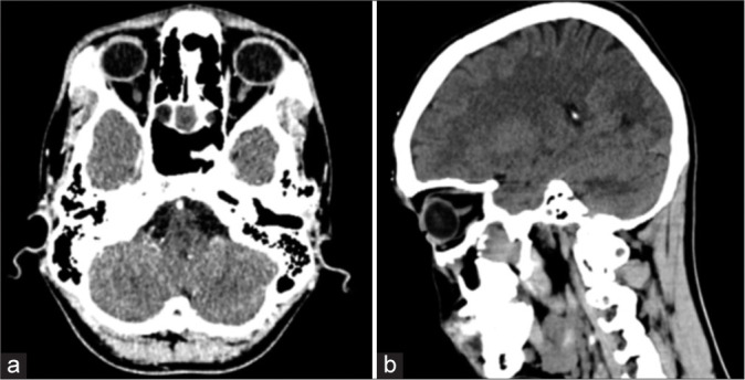
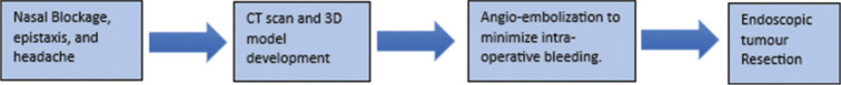
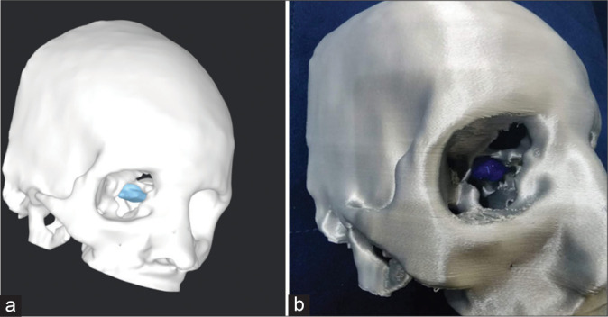
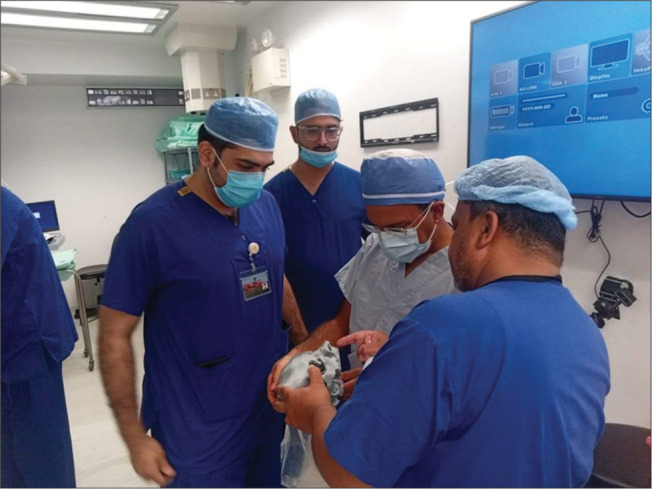
Our case is of a 19-year-old male with a 2-month history of recurrent epistaxis, nasal blockage, and headache. Based on the computed tomography scan and the clinical presentation, the patient was diagnosed with juvenile nasopharyngeal angiofibroma. The patient underwent angioembolization of the tumor followed by endoscopic surgical resection. The patient remained stable postoperatively and demonstrated a good recovery in the follow-up visit with no signs of cranial deficits. This case report highlights the use of a patient-specific 3D-printed biomodel to visualize this rare tumor of the nasopharynx. The benefits of using the model in surgical planning, patient education, and resident training are reported. We found that the ability to visualize the tumor on a tangible model, viewing its actual size in relation to the adjacent anatomy and all the structures associated with it, greatly enhances the surgeon's capacity to tackle such a difficult tumor endoscopically.

CONCLUSION

Incorporating 3D-printed biomodels in surgical practice should result in improved outcomes for the patients.

摘要

背景

三维(3D)打印已在各个领域证明了其作用。最近,3D打印也已引入耳鼻喉科领域。鼻咽、鼻窦和前颅底解剖结构复杂。在手术过程中必须小心保护关键结构。因此,对于外科医生来说,对所涉及的复杂手术区域有出色的空间理解非常重要。

病例描述

我们的病例是一名19岁男性,有2个月反复鼻出血、鼻塞和头痛的病史。根据计算机断层扫描和临床表现,该患者被诊断为青少年鼻咽血管纤维瘤。患者先接受了肿瘤血管栓塞术,随后进行了内镜手术切除。患者术后情况稳定,随访时恢复良好,无颅神经缺损迹象。本病例报告强调了使用患者特异性3D打印生物模型来可视化这种罕见的鼻咽肿瘤。报告了在手术规划、患者教育和住院医师培训中使用该模型的益处。我们发现,能够在实体模型上可视化肿瘤,查看其相对于相邻解剖结构及与之相关的所有结构的实际大小,极大地增强了外科医生通过内镜处理此类困难肿瘤的能力。

结论

将3D打印生物模型纳入手术实践应能改善患者的治疗效果。

https://cdn.ncbi.nlm.nih.gov/pmc/blobs/e82f/10927217/2a09108d2f8e/SNI-15-44-g004.jpg
https://cdn.ncbi.nlm.nih.gov/pmc/blobs/e82f/10927217/d027b2c5586d/SNI-15-44-g001.jpg
https://cdn.ncbi.nlm.nih.gov/pmc/blobs/e82f/10927217/44a8fe16a61b/SNI-15-44-g002.jpg
https://cdn.ncbi.nlm.nih.gov/pmc/blobs/e82f/10927217/d044c10fd458/SNI-15-44-g003.jpg
https://cdn.ncbi.nlm.nih.gov/pmc/blobs/e82f/10927217/2a09108d2f8e/SNI-15-44-g004.jpg

相似文献

1
Successful use of a patient specific 3D-printed biomodel as surgical guide for excision of juvenile nasopharyngeal angiofibroma extending to skull base: A case report.成功使用患者特异性3D打印生物模型作为手术指南切除延伸至颅底的青少年鼻咽血管纤维瘤:一例报告。
Surg Neurol Int. 2024 Feb 16;15:44. doi: 10.25259/SNI_743_2023. eCollection 2024.
2
Assessment of a Patient-Specific, 3-Dimensionally Printed Endoscopic Sinus and Skull Base Surgical Model.患者特定的三维打印内镜鼻窦和颅底手术模型评估。
JAMA Otolaryngol Head Neck Surg. 2018 Jul 1;144(7):574-579. doi: 10.1001/jamaoto.2018.0473.
3
Endoscopic graduated multiangle, multicorridor resection of juvenile nasopharyngeal angiofibroma: an individualized, tailored, multicorridor skull base approach.鼻内镜下分次多角度、多通道切除青少年鼻咽血管纤维瘤:一种个体化、定制化的多通道颅底入路
J Neurosurg. 2016 May;124(5):1328-38. doi: 10.3171/2014.12.JNS141696. Epub 2015 Nov 13.
4
A novel three-dimensional-printed paranasal sinus-skull base anatomical model.一种新型的三维打印的鼻窦-颅底解剖模型。
Eur Arch Otorhinolaryngol. 2018 Aug;275(8):2045-2049. doi: 10.1007/s00405-018-5051-z. Epub 2018 Jun 29.
5
Use of patient-specific 3D printed models in pre-operative counseling for pediatric skull base surgery.在小儿颅底外科手术的术前咨询中使用患者特异性 3D 打印模型。
Int J Pediatr Otorhinolaryngol. 2023 Aug;171:111655. doi: 10.1016/j.ijporl.2023.111655. Epub 2023 Jul 13.
6
Expanded endonasal endoscopic approach for resection of a juvenile nasopharyngeal angiofibroma with skull base involvement.扩大经鼻内镜入路切除颅底受累的青少年鼻咽血管纤维瘤。
J Clin Neurosci. 2010 Nov;17(11):1423-7. doi: 10.1016/j.jocn.2010.04.013. Epub 2010 Aug 13.
7
Resection of a juvenile nasopharyngeal angiofibroma via a hybrid cranioendoscopic approach.经颅内镜联合入路切除青少年鼻咽血管纤维瘤
Neurosurg Focus Video. 2020 Apr 1;2(2):V17. doi: 10.3171/2020.4.FocusVid.19923. eCollection 2020 Apr.
8
Bulky Recurrent Juvenile Nasopharyngeal Angiofibroma.巨大复发性青少年鼻咽血管纤维瘤
J Craniofac Surg. 2023;34(5):e468-e470. doi: 10.1097/SCS.0000000000009307. Epub 2023 Apr 14.
9
Rare presentation of angiomatosis in the paranasal sinuses mimicking juvenile nasopharyngeal angiofibroma in a 16 year old male.16岁男性鼻窦血管瘤病罕见表现,酷似青少年鼻咽血管纤维瘤。
Radiol Case Rep. 2022 Jul 29;17(10):3527-3534. doi: 10.1016/j.radcr.2022.07.031. eCollection 2022 Oct.
10
The utility of 3-dimensional-printed models for skull base meningioma surgery.三维打印模型在颅底脑膜瘤手术中的应用
Ann Transl Med. 2020 Mar;8(6):370. doi: 10.21037/atm.2020.02.28.

引用本文的文献

1
A Scoping Review of Pakistani Healthcare Simulation: Insights for Lower-Middle-Income Countries.巴基斯坦医疗模拟的范围综述:对中低收入国家的启示
Cureus. 2024 Dec 27;16(12):e76485. doi: 10.7759/cureus.76485. eCollection 2024 Dec.
2
Can high-fidelity 3D models be a good alternative for cadaveric materials in skill assessment for endoscopic sinus surgery? A comparison study in assessment for surgical performance in 3D models and cadavers.高保真3D模型能否成为尸体材料在内镜鼻窦手术技能评估中的良好替代物?一项关于3D模型和尸体手术表现评估的比较研究。
Front Med (Lausanne). 2024 Oct 17;11:1301511. doi: 10.3389/fmed.2024.1301511. eCollection 2024.

本文引用的文献

1
Use of patient-specific 3D printed models in pre-operative counseling for pediatric skull base surgery.在小儿颅底外科手术的术前咨询中使用患者特异性 3D 打印模型。
Int J Pediatr Otorhinolaryngol. 2023 Aug;171:111655. doi: 10.1016/j.ijporl.2023.111655. Epub 2023 Jul 13.
2
Current approach of juvenile nasopharyngeal angiofibroma: a case series.青少年鼻咽血管纤维瘤的当前治疗方法:病例系列。
Rom J Morphol Embryol. 2022 Jan-Mar;63(1):105-111. doi: 10.47162/RJME.63.1.10.
3
A case report detailing the use of 3D printing technology in surgical planning and decision making in ENT surgery-an axial 3D first in Northern Ireland.
一份病例报告详细介绍了3D打印技术在耳鼻喉科手术的手术规划和决策中的应用——这是北爱尔兰首例轴向3D打印应用。
Int J Surg Case Rep. 2021 Oct;87:106407. doi: 10.1016/j.ijscr.2021.106407. Epub 2021 Sep 15.
4
Patient-specific 3-dimensionally printed models for neurosurgical planning and education.用于神经外科规划和教育的患者特异性三维打印模型。
Neurosurg Focus. 2019 Dec 1;47(6):E12. doi: 10.3171/2019.9.FOCUS19511.
5
Three-Dimensional Printing: Current Use in Rhinology and Endoscopic Skull Base Surgery.三维打印:目前在鼻科学和内镜颅底手术中的应用
Am J Rhinol Allergy. 2019 Nov;33(6):770-781. doi: 10.1177/1945892419866319. Epub 2019 Aug 5.
6
A small 3D-printing model of macroadenomas for endoscopic endonasal surgery.用于经鼻内镜手术的大型腺瘤的小型 3D 打印模型。
Pituitary. 2019 Feb;22(1):46-53. doi: 10.1007/s11102-018-0927-x.
7
3D printing and intraoperative neuronavigation tailoring for skull base reconstruction after extended endoscopic endonasal surgery: proof of concept.3D 打印与术中神经导航技术在扩大经鼻内镜颅底手术后颅底重建中的应用:概念验证。
J Neurosurg. 2019 Jan 1;130(1):248-255. doi: 10.3171/2017.9.JNS171253.
8
Patient Education for Endoscopic Sinus Surgery: Preliminary Experience Using 3D-Printed Clinical Imaging Data.内镜鼻窦手术的患者教育:使用3D打印临床影像数据的初步经验
J Funct Biomater. 2017 Apr 7;8(2):13. doi: 10.3390/jfb8020013.
9
3D printed renal cancer models derived from MRI data: application in pre-surgical planning.基于 MRI 数据的 3D 打印肾癌模型:在术前规划中的应用。
Abdom Radiol (NY). 2017 May;42(5):1501-1509. doi: 10.1007/s00261-016-1022-2.
10
Endoscopic skull base training using 3D printed models with pre-existing pathology.使用带有既往病变的3D打印模型进行内镜颅底手术训练。
Eur Arch Otorhinolaryngol. 2015 Mar;272(3):753-7. doi: 10.1007/s00405-014-3300-3. Epub 2014 Oct 8.