Badge Mohanish, Kapoor Poonam Malhotra, Thiruselvan T, Francis Jijo
Department of Cardiac Anaesthesia and Critical Care, All India Institute of Medical Sciences, New Delhi, Delhi, India.
J Clin Imaging Sci. 2024 Feb 29;14:6. doi: 10.25259/JCIS_136_2023. eCollection 2024.
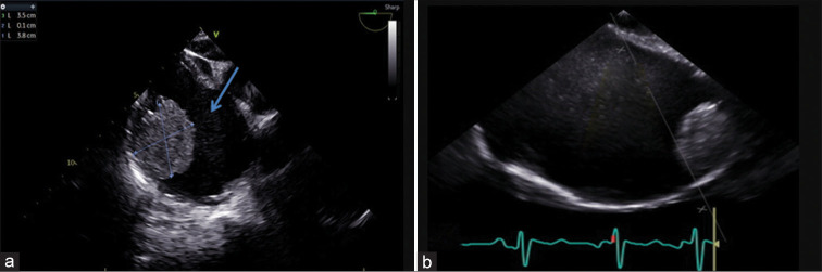
Cardiac masses are a significant cause of patient morbidity and mortality by virtue of their symptoms and surgical removal. Preoperative diagnosis of a cardiac mass is usually based on clinical correlation and transthoracic echocardiography findings. Myxomas are the most common benign cardiac tumors, commonly occurring in the left atrium attached to the interatrial septum near the fossa ovalis. Although, at times atypical location and unusual morphology may pose a diagnostic dilemma with 2D echocardiography. 3D echocardiography with its multifaceted advantages, including multiplanar cropping abilities and superior imaging quality can help distinguish between a clot and a myxoma.
心脏肿物因其症状及手术切除情况,是导致患者发病和死亡的重要原因。心脏肿物的术前诊断通常基于临床关联及经胸超声心动图检查结果。黏液瘤是最常见的良性心脏肿瘤,通常发生于左心房,附着于卵圆窝附近的房间隔。然而,有时非典型位置和异常形态可能会给二维超声心动图带来诊断难题。三维超声心动图具有多方面优势,包括多平面裁剪能力和卓越的成像质量,有助于区分血栓和黏液瘤。