• 文献检索
  • 文档翻译
  • 深度研究
  • 学术资讯
  • Suppr Zotero 插件Zotero 插件
  • 邀请有礼
  • 套餐&价格
  • 历史记录
应用&插件
Suppr Zotero 插件Zotero 插件浏览器插件Mac 客户端Windows 客户端微信小程序
定价
高级版会员购买积分包购买API积分包
服务
文献检索文档翻译深度研究API 文档MCP 服务
关于我们
关于 Suppr公司介绍联系我们用户协议隐私条款
关注我们

Suppr 超能文献

核心技术专利:CN118964589B侵权必究
粤ICP备2023148730 号-1Suppr @ 2026

文献检索

告别复杂PubMed语法,用中文像聊天一样搜索,搜遍4000万医学文献。AI智能推荐,让科研检索更轻松。

立即免费搜索

文件翻译

保留排版,准确专业,支持PDF/Word/PPT等文件格式,支持 12+语言互译。

免费翻译文档

深度研究

AI帮你快速写综述,25分钟生成高质量综述,智能提取关键信息,辅助科研写作。

立即免费体验

心脏肿瘤:超声评估

Cardiac tumors: echo assessment.

作者信息

Mankad Rekha, Herrmann Joerg

机构信息

Department of Cardiovascular Diseases, Mayo Clinic, Rochester, Minnesota, USA.

Department of Cardiovascular Diseases, Mayo Clinic, Rochester, Minnesota, USA

出版信息

Echo Res Pract. 2016 Dec;3(4):R65-R77. doi: 10.1530/ERP-16-0035. Epub 2016 Sep 6.

DOI:10.1530/ERP-16-0035
PMID:27600455
原文链接:https://pmc.ncbi.nlm.nih.gov/articles/PMC5292983/
Abstract

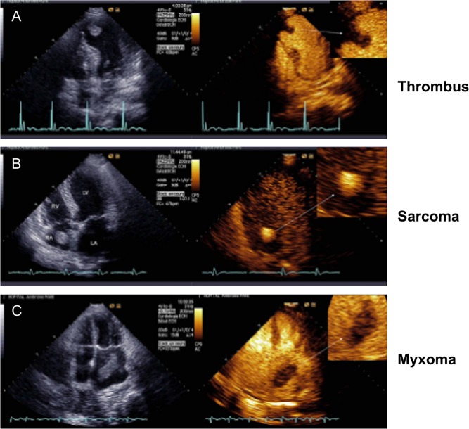
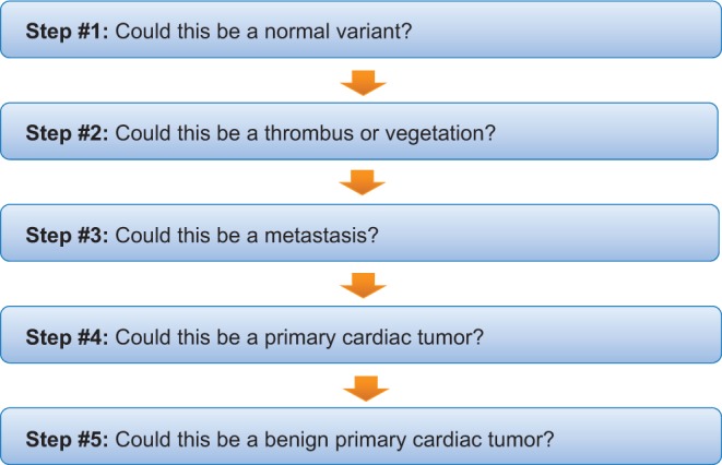
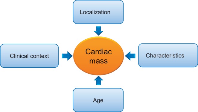
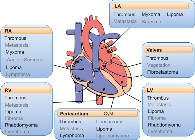
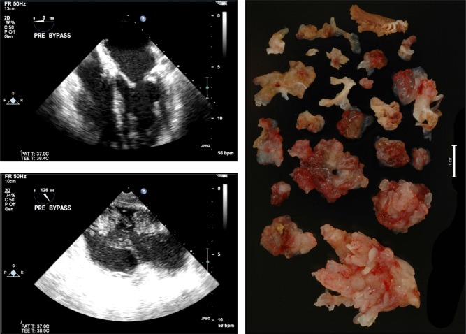
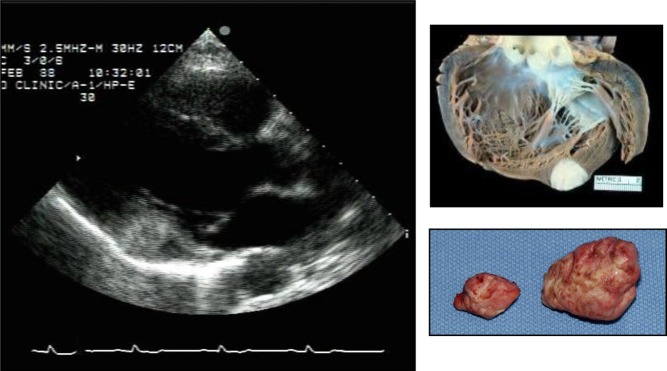
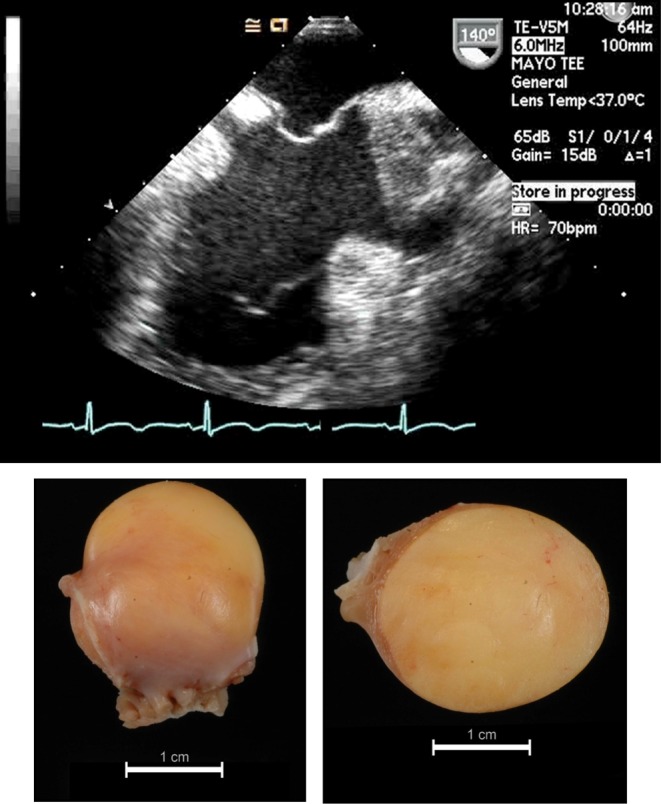
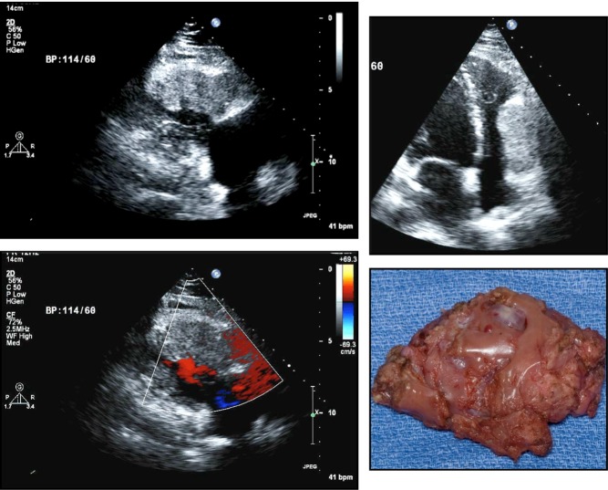
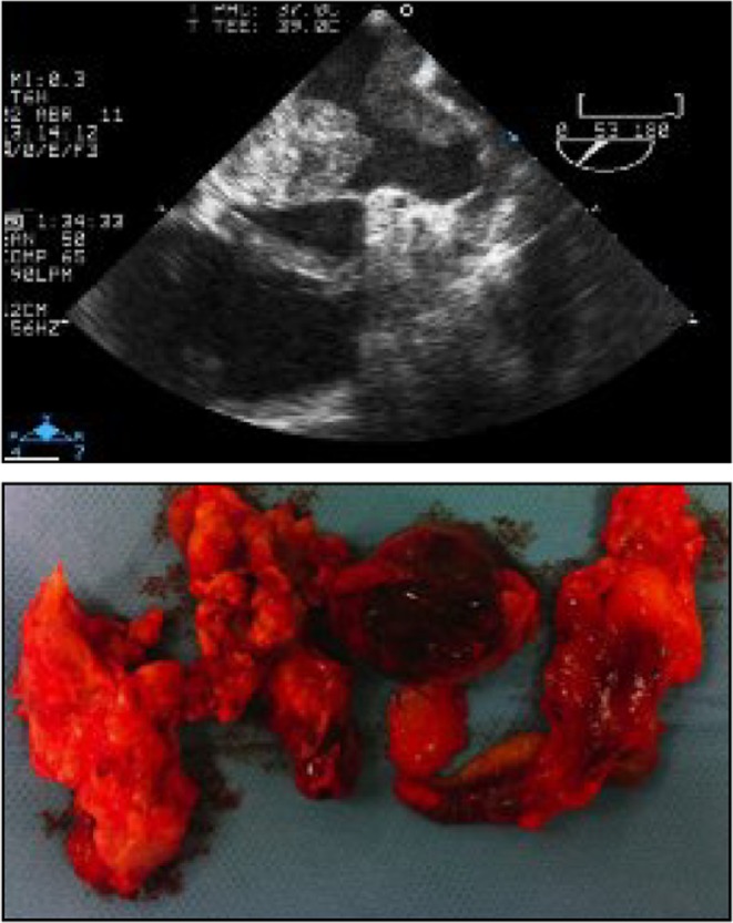
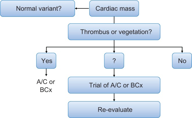
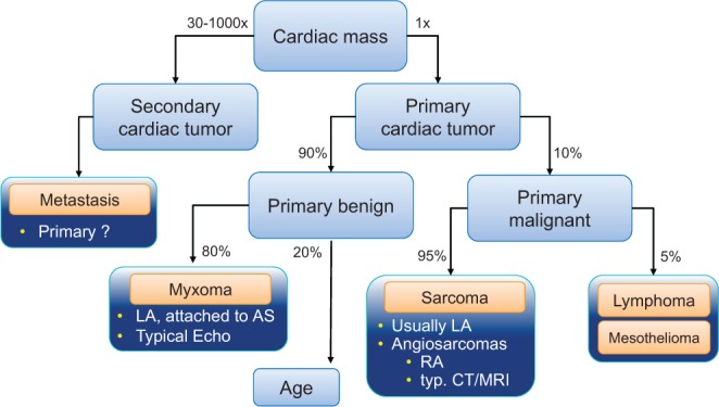
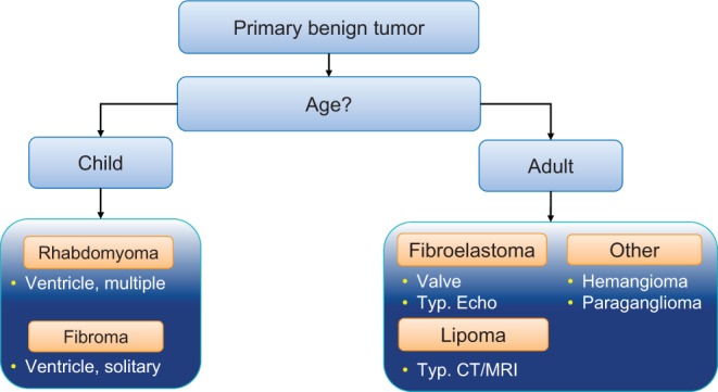
Cardiac tumors are exceedingly rare (0.001-0.03% in most autopsy series). They can be present anywhere within the heart and can be attached to any surface or be embedded in the myocardium or pericardial space. Signs and symptoms are nonspecific and highly variable related to the localization, size and composition of the cardiac mass. Echocardiography, typically performed for another indication, may be the first imaging modality alerting the clinician to the presence of a cardiac mass. Although echocardiography cannot give the histopathology, certain imaging features and adjunctive tools such as contrast imaging may aid in the differential diagnosis as do the adjunctive clinical data and the following principles: (1) thrombus or vegetations are the most likely etiology, (2) cardiac tumors are mostly secondary and (3) primary cardiac tumors are mostly benign. Although the finding of a cardiac mass on echocardiography may generate confusion, a stepwise approach may serve well practically. Herein, we will review such an approach and the role of echocardiography in the assessment of cardiac masses.

摘要

心脏肿瘤极为罕见(在大多数尸检系列中占0.001%-0.03%)。它们可出现在心脏内的任何部位,可附着于任何表面,或嵌入心肌或心包腔。体征和症状是非特异性的,且因心脏肿物的位置、大小和组成不同而有很大差异。超声心动图通常因其他指征而进行检查,可能是提醒临床医生存在心脏肿物的首个影像学检查手段。虽然超声心动图无法提供组织病理学结果,但某些影像学特征以及诸如造影成像等辅助工具,与辅助临床数据及以下原则一样,有助于鉴别诊断:(1)血栓或赘生物是最可能的病因,(2)心脏肿瘤大多是继发性的,(3)原发性心脏肿瘤大多是良性的。虽然超声心动图发现心脏肿物可能会造成混淆,但采用逐步分析的方法在实际应用中可能效果良好。在此,我们将回顾这种方法以及超声心动图在评估心脏肿物中的作用。

https://cdn.ncbi.nlm.nih.gov/pmc/blobs/ed80/5292983/abfb4f62b837/erp-3-R65-g019.jpg
https://cdn.ncbi.nlm.nih.gov/pmc/blobs/ed80/5292983/864a3378f865/erp-3-R65-g001.jpg
https://cdn.ncbi.nlm.nih.gov/pmc/blobs/ed80/5292983/c9401bc9c9f4/erp-3-R65-g002.jpg
https://cdn.ncbi.nlm.nih.gov/pmc/blobs/ed80/5292983/ebc6750243fc/erp-3-R65-g003.jpg
https://cdn.ncbi.nlm.nih.gov/pmc/blobs/ed80/5292983/1cc0f99a2fe0/erp-3-R65-g004.jpg
https://cdn.ncbi.nlm.nih.gov/pmc/blobs/ed80/5292983/33630e0c8eb5/erp-3-R65-g005.jpg
https://cdn.ncbi.nlm.nih.gov/pmc/blobs/ed80/5292983/d43cf8b7200f/erp-3-R65-g006.jpg
https://cdn.ncbi.nlm.nih.gov/pmc/blobs/ed80/5292983/d0ce350033ab/erp-3-R65-g007.jpg
https://cdn.ncbi.nlm.nih.gov/pmc/blobs/ed80/5292983/119f6f5f35fc/erp-3-R65-g008.jpg
https://cdn.ncbi.nlm.nih.gov/pmc/blobs/ed80/5292983/4602ea430f79/erp-3-R65-g009.jpg
https://cdn.ncbi.nlm.nih.gov/pmc/blobs/ed80/5292983/b9d1bbec9505/erp-3-R65-g010.jpg
https://cdn.ncbi.nlm.nih.gov/pmc/blobs/ed80/5292983/9f4eae44eff8/erp-3-R65-g011.jpg
https://cdn.ncbi.nlm.nih.gov/pmc/blobs/ed80/5292983/07938c9d1a42/erp-3-R65-g012.jpg
https://cdn.ncbi.nlm.nih.gov/pmc/blobs/ed80/5292983/7aadc4f561a5/erp-3-R65-g013.jpg
https://cdn.ncbi.nlm.nih.gov/pmc/blobs/ed80/5292983/361106523180/erp-3-R65-g014.jpg
https://cdn.ncbi.nlm.nih.gov/pmc/blobs/ed80/5292983/509cff527b3a/erp-3-R65-g015.jpg
https://cdn.ncbi.nlm.nih.gov/pmc/blobs/ed80/5292983/1bd40f1f023a/erp-3-R65-g016.jpg
https://cdn.ncbi.nlm.nih.gov/pmc/blobs/ed80/5292983/438b21f52aed/erp-3-R65-g017.jpg
https://cdn.ncbi.nlm.nih.gov/pmc/blobs/ed80/5292983/9db26f0529fb/erp-3-R65-g018.jpg
https://cdn.ncbi.nlm.nih.gov/pmc/blobs/ed80/5292983/abfb4f62b837/erp-3-R65-g019.jpg

相似文献

1
Cardiac tumors: echo assessment.心脏肿瘤:超声评估
Echo Res Pract. 2016 Dec;3(4):R65-R77. doi: 10.1530/ERP-16-0035. Epub 2016 Sep 6.
2
Usefulness of contrast perfusion echocardiography for differential diagnosis of cardiac masses.对比剂灌注超声心动图在心脏肿物鉴别诊断中的应用价值
Ultrasound Med Biol. 2015 Sep;41(9):2382-90. doi: 10.1016/j.ultrasmedbio.2015.05.010. Epub 2015 Jun 16.
3
CT and MR imaging of benign primary cardiac neoplasms with echocardiographic correlation.良性原发性心脏肿瘤的CT和MR成像与超声心动图相关性研究
Radiographics. 2000 Sep-Oct;20(5):1303-19. doi: 10.1148/radiographics.20.5.g00se121303.
4
Evaluation and Management of Cardiac Tumors.心脏肿瘤的评估与管理
Curr Treat Options Cardiovasc Med. 2018 Mar 20;20(4):29. doi: 10.1007/s11936-018-0625-z.
5
Comparison of echo and MRI in the imaging evaluation of intracardiac masses.超声心动图与磁共振成像在心脏内肿块成像评估中的比较。
Cardiovasc Intervent Radiol. 2004 Sep-Oct;27(5):459-69. doi: 10.1007/s00270-004-0123-4. Epub 2004 Jun 16.
6
Cardiac Masses: The Role of Cardiovascular Imaging in the Differential Diagnosis.心脏肿物:心血管成像在鉴别诊断中的作用
Diagnostics (Basel). 2020 Dec 14;10(12):1088. doi: 10.3390/diagnostics10121088.
7
Biatrial thrombi resembling myxoma regressed after prolonged anticoagulation in a patient with mitral stenosis: a case report.二尖瓣狭窄患者经长期抗凝治疗后,类似黏液瘤的双房血栓消退:一例病例报告
J Med Case Rep. 2016 Aug 10;10(1):221. doi: 10.1186/s13256-016-1018-0.
8
[Improved structure identification with tissue Doppler echocardiography].[利用组织多普勒超声心动图改进结构识别]
Herz. 1998 Dec;23(8):499-505. doi: 10.1007/BF03043757.
9
A right atrial mass and a pseudomass.一个右心房肿块和一个假肿块。
Echocardiography. 2005 May;22(5):441-4. doi: 10.1111/j.1540-8175.2005.04049.x.
10
Echocardiographic Evaluation of Cardiac Masses.超声心动图评估心脏肿块。
Curr Cardiol Rep. 2023 Oct;25(10):1281-1290. doi: 10.1007/s11886-023-01945-z. Epub 2023 Sep 20.

引用本文的文献

1
Blunt Cardiac Injury Resulting in Avulsion of Septal Myocardium Without Ventricular Septal Defect.钝性心脏损伤导致间隔心肌撕裂但无室间隔缺损。
Cureus. 2025 Jun 10;17(6):e85722. doi: 10.7759/cureus.85722. eCollection 2025 Jun.
2
Diagnosis and treatment of cardiac tumors.心脏肿瘤的诊断与治疗。
Med Oncol. 2025 Mar 17;42(4):110. doi: 10.1007/s12032-025-02661-0.
3
Cardiac myxoma: a comprehensive review.心脏黏液瘤:全面综述。

本文引用的文献

1
Echocardiographic Features of Cardiac Angiosarcomas: The Mayo Clinic Experience (1976-2013).心脏血管肉瘤的超声心动图特征:梅奥诊所经验(1976 - 2013年)
Echocardiography. 2016 Feb;33(2):186-92. doi: 10.1111/echo.13060. Epub 2015 Oct 13.
2
Cardiac Myxomas: A 50-Year Experience With Resection and Analysis of Risk Factors for Recurrence.心脏黏液瘤:50年手术切除经验及复发危险因素分析
Ann Thorac Surg. 2015 Aug;100(2):495-500. doi: 10.1016/j.athoracsur.2015.03.007. Epub 2015 Jun 9.
3
Prognostic and Bioepidemiologic Implications of Papillary Fibroelastomas.
J Cardiothorac Surg. 2025 Mar 13;20(1):151. doi: 10.1186/s13019-024-03333-2.
4
Diagnostic Algorithm Using Multimodal Imaging for the Differential Diagnosis of Intra-Cardiac Masses.使用多模态成像进行心内肿物鉴别诊断的诊断算法
J Clin Med. 2025 Jan 15;14(2):508. doi: 10.3390/jcm14020508.
5
Could Pulsed Wave Tissue Doppler Imaging Solve the Diagnostic Dilemma of Right Atrial Masses and Pseudomasses? A Case Series and Literature Review.脉冲波组织多普勒成像能否解决右心房肿块和假肿块的诊断难题?病例系列研究与文献综述
J Clin Med. 2024 Dec 27;14(1):86. doi: 10.3390/jcm14010086.
6
A massive cardiac lipoma in the right atrium with multimodality imaging.一例右心房巨大心脏脂肪瘤的多模态影像学表现
J Cardiothorac Surg. 2024 Dec 19;19(1):646. doi: 10.1186/s13019-024-03162-3.
7
A Rare Case of Inflammatory Cardiac Mass in the Young: Diagnostic Uncertainty and Therapeutic Challenges.青年炎性心脏肿物一例:诊断的不确定性与治疗挑战
Cureus. 2024 Oct 31;16(10):e72791. doi: 10.7759/cureus.72791. eCollection 2024 Oct.
8
Total resection via right mini-thoracotomy for left atrial myxoma in juvenile Carney complex: a case report.通过右胸小切口对患有卡尼综合征的青少年进行左心房黏液瘤全切除术:病例报告
Gen Thorac Cardiovasc Surg Cases. 2024 Oct 29;3(1):48. doi: 10.1186/s44215-024-00173-3.
9
Multimodal Imaging Approach to Atrial Myxoma to Optimize Surgical Management.用于优化手术管理的心房黏液瘤多模态成像方法
Cureus. 2024 Aug 12;16(8):e66719. doi: 10.7759/cureus.66719. eCollection 2024 Aug.
10
Mantle Cell Lymphoma With Cardiac Involvement Presenting as Complete Heart Block.表现为完全性心脏传导阻滞的合并心脏受累的套细胞淋巴瘤。
JACC Case Rep. 2024 Aug 7;29(15):102416. doi: 10.1016/j.jaccas.2024.102416.
乳头状弹性纤维瘤的预后和生物流行病学意义。
J Am Coll Cardiol. 2015 Jun 9;65(22):2420-9. doi: 10.1016/j.jacc.2015.03.569.
4
Aortic valve myxoma at the extreme age: a review of literature.高龄患者的主动脉瓣黏液瘤:文献综述
BMJ Case Rep. 2014 Mar 18;2014:bcr2013202689. doi: 10.1136/bcr-2013-202689.
5
Tumors metastatic to the heart.转移性心脏肿瘤。
Circulation. 2013 Oct 15;128(16):1790-4. doi: 10.1161/CIRCULATIONAHA.112.000790.
6
Malignant primary cardiac tumours.原发性恶性心脏肿瘤。
Interact Cardiovasc Thorac Surg. 2012 Dec;15(6):1004-6. doi: 10.1093/icvts/ivs350. Epub 2012 Aug 24.
7
Hemoptysis as the presenting symptom in cardiac sarcoma.咯血作为心脏肉瘤的首发症状。
Rev Port Cardiol. 2012 Jun;31(6):463-4. doi: 10.1016/j.repc.2012.04.005.
8
Real time three-dimensional echocardiography evaluation of intracardiac masses.心内肿物的实时三维超声心动图评估
Echocardiography. 2012 Feb;29(2):207-19. doi: 10.1111/j.1540-8175.2011.01627.x.
9
Cardiac tumours: diagnosis and management.心脏肿瘤:诊断与管理
Heart. 2011 Jan;97(2):151-60. doi: 10.1136/hrt.2009.186320.
10
Images in cardiovascular medicine. Primary cardiac lymphoma: a rare case of pulmonary tumor embolism.心血管医学影像。原发性心脏淋巴瘤:一例罕见的肺肿瘤栓塞病例。
Circulation. 2010 May 25;121(20):2249-50. doi: 10.1161/CIRCULATIONAHA.109.863126.