Hussein Nancy, Meade Josephine, Pandit Hemant, Jones Elena, El-Gendy Reem
Division of Oral Biology, School of Dentistry, University of Leeds, Leeds LS9 7TF, UK.
Department of Oral Medicine and Periodontology, Faculty of Dentistry, Mansoura University, Mansoura 35516, Egypt.
Int J Mol Sci. 2024 Mar 1;25(5):2851. doi: 10.3390/ijms25052851.
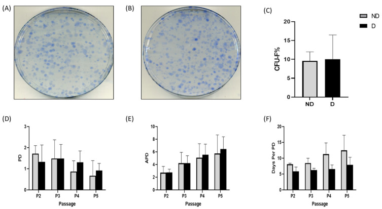
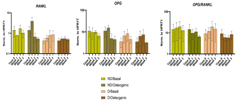
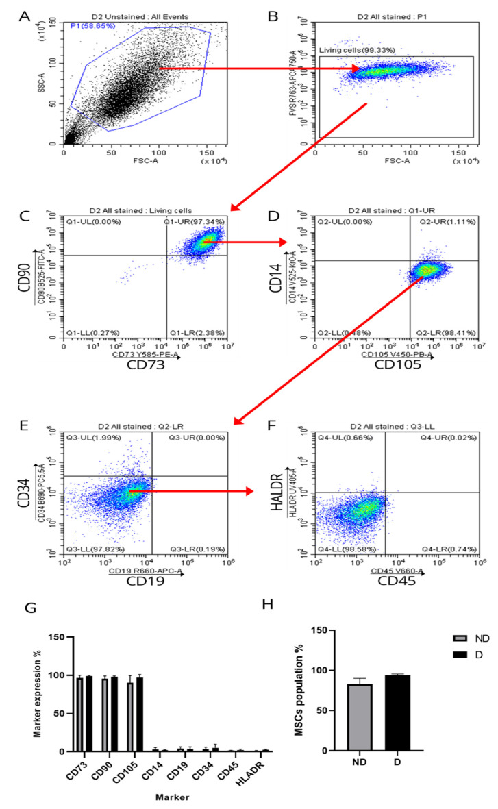
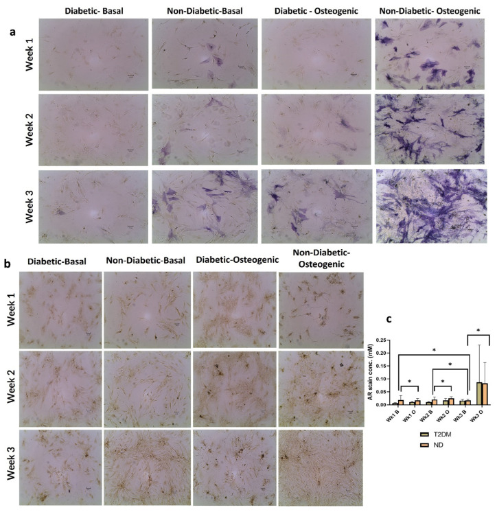
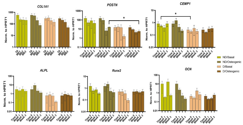
Type 2 diabetes mellitus (T2DM) represents a significant health problem globally and is linked to a number of complications such as cardiovascular disease, bone fragility and periodontitis. Autologous bone marrow mesenchymal stem cells (BM-MSCs) are a promising therapeutic approach for bone and periodontal regeneration; however, the effect of T2DM on the expression of osteogenic and periodontal markers in BM-MSCs is not fully established. Furthermore, the effect of the presence of comorbidities such as diabetes and osteoarthritis on BM-MSCs is also yet to be investigated. In the present study, BM-MSCs were isolated from osteoarthritic knee joints of diabetic and nondiabetic donors. Both cell groups were compared for their clonogenicity, proliferation rates, MSC enumeration and expression of surface markers. Formation of calcified deposits and expression of osteogenic and periodontal markers were assessed after 1, 2 and 3 weeks of basal and osteogenic culture. Diabetic and nondiabetic BM-MSCs showed similar clonogenic and growth potentials along with comparable numbers of MSCs. However, diabetic BM-MSCs displayed lower expression of periostin (POSTN) and cementum protein 1 (CEMP-1) at Wk3 osteogenic and Wk1 basal cultures, respectively. BM-MSCs from T2DM patients might be suitable candidates for stem cell-based therapeutics. However, further investigations into these cells' behaviours in vitro and in vivo under inflammatory environments and hyperglycaemic conditions are still required.
2型糖尿病(T2DM)是全球范围内一个重大的健康问题,与多种并发症相关,如心血管疾病、骨质脆弱和牙周炎。自体骨髓间充质干细胞(BM-MSCs)是一种有前景的骨和牙周组织再生治疗方法;然而,T2DM对BM-MSCs中成骨和牙周标志物表达的影响尚未完全明确。此外,糖尿病和骨关节炎等合并症对BM-MSCs的影响也有待研究。在本研究中,从糖尿病和非糖尿病供体的骨关节炎膝关节中分离出BM-MSCs。比较了两组细胞的克隆形成能力、增殖率、MSC计数和表面标志物的表达。在基础培养和成骨培养1、2和3周后,评估钙化沉积物的形成以及成骨和牙周标志物的表达。糖尿病和非糖尿病BM-MSCs表现出相似的克隆形成和生长潜力,以及相当数量的MSC。然而,糖尿病BM-MSCs在成骨培养第3周和基础培养第1周时,分别显示出较低的骨膜蛋白(POSTN)和牙骨质蛋白1(CEMP-1)表达。来自T2DM患者的BM-MSCs可能是基于干细胞治疗的合适候选者。然而,仍需要进一步研究这些细胞在炎症环境和高血糖条件下的体外和体内行为。