Dong Yiming, Wang Jianfeng, Yang Chenteng, Bao Junxia, Liu Xia, Chen Hao, Zhang Xiaojing, Shi Weibo, Zhang Lihua, Qi Qian, Li Yingmin, Wang Songjun, Ma Rufei, Cong Bin, Zhang Guozhong
Hebei Key Laboratory of Forensic Medicine, Collaborative Innovation Center of Forensic Medical Molecular Identification, College of Forensic Medicine, Hebei Medical University, Shijiazhuang 050017, China.
Hebei Province Laboratory of Experimental Animal, Shijiazhuang 050017, China.
Int J Mol Sci. 2024 Mar 3;25(5):2941. doi: 10.3390/ijms25052941.
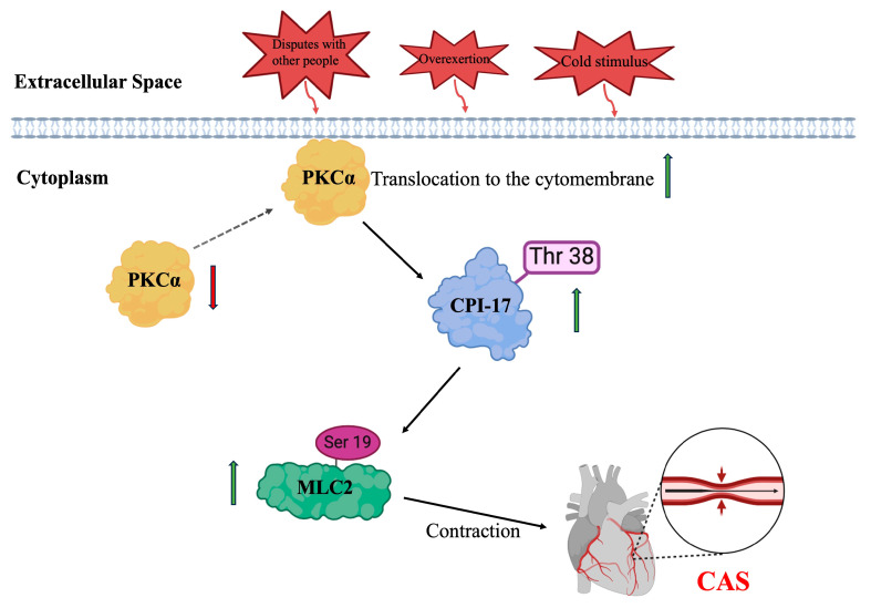
Coronary artery spasm (CAS) plays an important role in the pathogeneses of various ischemic heart diseases and has gradually become a common cause of life-threatening arrhythmia. The specific molecular mechanism of CAS has not been fully elucidated, nor are there any specific diagnostic markers for the condition. Therefore, this study aimed to examine the specific molecular mechanism underlying CAS, and screen for potential diagnostic markers. To this end, we successfully constructed a rat CAS model and achieved in vitro culture of a human coronary-artery smooth-muscle cell (hCASMC) contraction model. Possible molecular mechanisms by which protein kinase C (PKC) regulated CAS through the C kinase-potentiated protein phosphatase 1 inhibitor of 17 kDa (CPI-17)/myosin II regulatory light chain (MLC2) pathway were studied in vivo and in vitro to screen for potential molecular markers of CAS. We performed hematoxylin and eosin staining, myocardial zymogram, and transmission electron microscopy to determine myocardial and coronary artery injury in CAS rats. Then, using immunohistochemical staining, immunofluorescence staining, and Western blotting, we further demonstrated a potential molecular mechanism by which PKC regulated CAS via the CPI-17/MLC2 pathway. The results showed that membrane translocation of PKCα occurred in the coronary arteries of CAS rats. CPI-17/MLC2 signaling was observably activated in coronary arteries undergoing CAS. In addition, in vitro treatment of hCASMCs with angiotensin II (Ang II) increased PKCα membrane translocation while consistently activating CPI-17/MLC2 signaling. Conversely, GF-109203X and calphostin C, specific inhibitors of PKC, inactivated CPI-17/MLC2 signaling. We also collected the coronary artery tissues from deceased subjects suspected to have died of CAS and measured their levels of phosphorylated CPI-17 (p-CPI-17) and MLC2 (p-MLC2). Immunohistochemical staining was positive for p-CPI-17 and p-MLC2 in the tissues of these subjects. These findings suggest that PKCα induced CAS through the CPI-17/MLC2 pathway; therefore, p-CPI-17 and p-MLC2 could be used as potential markers for CAS. Our data provide novel evidence that therapeutic strategies against PKC or CPI-17/MLC2 signaling might be promising in the treatment of CAS.
冠状动脉痉挛(CAS)在各种缺血性心脏病的发病机制中起重要作用,并逐渐成为危及生命的心律失常的常见原因。CAS的具体分子机制尚未完全阐明,也没有针对该病症的特异性诊断标志物。因此,本研究旨在探讨CAS潜在的分子机制,并筛选潜在的诊断标志物。为此,我们成功构建了大鼠CAS模型,并实现了人冠状动脉平滑肌细胞(hCASMC)收缩模型的体外培养。在体内和体外研究了蛋白激酶C(PKC)通过17 kDa的C激酶增强蛋白磷酸酶1抑制剂(CPI-17)/肌球蛋白II调节轻链(MLC2)途径调节CAS的可能分子机制,以筛选CAS潜在的分子标志物。我们进行了苏木精-伊红染色、心肌酶谱分析和透射电子显微镜检查,以确定CAS大鼠的心肌和冠状动脉损伤。然后,通过免疫组织化学染色、免疫荧光染色和蛋白质印迹法,我们进一步证实了PKC通过CPI-17/MLC2途径调节CAS的潜在分子机制。结果显示,PKCα在CAS大鼠冠状动脉中发生膜转位。在发生CAS的冠状动脉中,CPI-17/MLC2信号明显激活。此外,用血管紧张素II(Ang II)体外处理hCASMC可增加PKCα膜转位,同时持续激活CPI-17/MLC2信号。相反,PKC的特异性抑制剂GF-109203X和钙泊三醇C可使CPI-17/MLC2信号失活。我们还收集了疑似死于CAS的死者的冠状动脉组织,并检测了其磷酸化CPI-17(p-CPI-17)和MLC2(p-MLC2)的水平。这些受试者组织中的p-CPI-17和p-MLC2免疫组织化学染色呈阳性。这些发现表明,PKCα通过CPI-17/MLC2途径诱导CAS;因此,p-CPI-17和p-MLC2可作为CAS的潜在标志物。我们的数据提供了新的证据,表明针对PKC或CPI-17/MLC2信号的治疗策略在CAS治疗中可能具有前景。