Department of Anesthesiology, The First Medical Center of Chinese PLA General Hospital, Beijing, China.
Department of Anesthesiology, The Sixth Medical Center of Chinese PLA General Hospital, Beijing, China.
CNS Neurosci Ther. 2024 Mar;30(3):e14675. doi: 10.1111/cns.14675.
General anesthesia has been used in surgical procedures for approximately 180 years, yet the precise mechanism of anesthetic drugs remains elusive. There is significant anatomical connectivity between the ventral tegmental area (VTA) and the prelimbic cortex (PrL). Projections from VTA dopaminergic neurons (VTA ) to the PrL play a role in the transition from sevoflurane anesthesia to arousal. It is still uncertain whether the prelimbic cortex pyramidal neuron (PrL ) and its projections to VTA (PrL -VTA) are involved in anesthesia-arousal regulation.
We employed chemogenetics and optogenetics to selectively manipulate neuronal activity in the PrL -VTA pathway. Electroencephalography spectra and burst-suppression ratios (BSR) were used to assess the depth of anesthesia. Furthermore, the loss or recovery of the righting reflex was monitored to indicate the induction or emergence time of general anesthesia. To elucidate the receptor mechanisms in the PrL -VTA projection's impact on anesthesia and arousal, we microinjected NMDA receptor antagonists (MK-801) or AMPA receptor antagonists (NBQX) into the VTA.
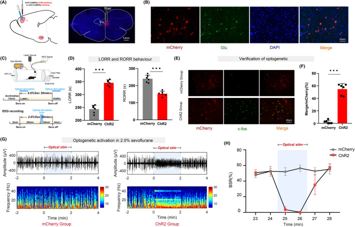
Our findings show that chemogenetic or optogenetic activation of PrL neurons prolonged anesthesia induction and promoted emergence. Additionally, chemogenetic activation of the PrL -VTA neural pathway delayed anesthesia induction and promoted anesthesia emergence. Likewise, optogenetic activation of the PrL -VTA projections extended the induction time and facilitated emergence from sevoflurane anesthesia. Moreover, antagonizing NMDA receptors in the VTA attenuates the delayed anesthesia induction and promotes emergence caused by activating the PrL -VTA projections.
This study demonstrates that PrL neurons and their projections to the VTA are involved in facilitating emergence from sevoflurane anesthesia, with the PrL -VTA pathway exerting its effects through the activation of NMDA receptors within the VTA.
全身麻醉已在外科手术中使用了大约 180 年,但麻醉药物的确切作用机制仍难以捉摸。腹侧被盖区(VTA)与额前皮质(PrL)之间存在显著的解剖连接。VTA 多巴胺能神经元(VTA)投射到 PrL 在七氟醚麻醉到觉醒的转变中起作用。尚不确定额前皮质锥体神经元(PrL)及其投射到 VTA(PrL-VTA)是否参与麻醉-觉醒调节。
我们采用化学遗传学和光遗传学选择性地操纵 PrL-VTA 通路中的神经元活动。脑电图频谱和爆发抑制比(BSR)用于评估麻醉深度。此外,监测翻正反射的丧失或恢复以指示全身麻醉的诱导或苏醒时间。为了阐明 PrL-VTA 投射中的受体机制对麻醉和觉醒的影响,我们将 NMDA 受体拮抗剂(MK-801)或 AMPA 受体拮抗剂(NBQX)微注射到 VTA 中。
我们的研究结果表明,化学遗传或光遗传激活 PrL 神经元可延长麻醉诱导时间并促进苏醒。此外,PrL-VTA 神经通路的化学遗传激活可延迟麻醉诱导并促进麻醉苏醒。同样,PrL-VTA 投射的光遗传激活可延长诱导时间并促进七氟醚麻醉的苏醒。此外,在 VTA 中拮抗 NMDA 受体可减弱激活 PrL-VTA 投射引起的麻醉诱导延迟和促进苏醒。
本研究表明,PrL 神经元及其投射到 VTA 参与促进七氟醚麻醉苏醒,PrL-VTA 通路通过 VTA 内 NMDA 受体的激活发挥作用。