Department of Experimental Psychology, University of Oxford, Tinsley Building, Mansfield Road, Oxford, OX1 3TA, UK.
Wellcome Centre for Integrative Neuroimaging (WIN), Centre for Functional MRI of the Brain (FMRIB, University of Oxford, Nuffield Department of Clinical Neurosciences, Level 6, West Wing, John Radcliffe Hospital, Oxford, OX3 9DU, UK.
Nat Commun. 2024 Mar 18;15(1):2426. doi: 10.1038/s41467-024-46275-y.
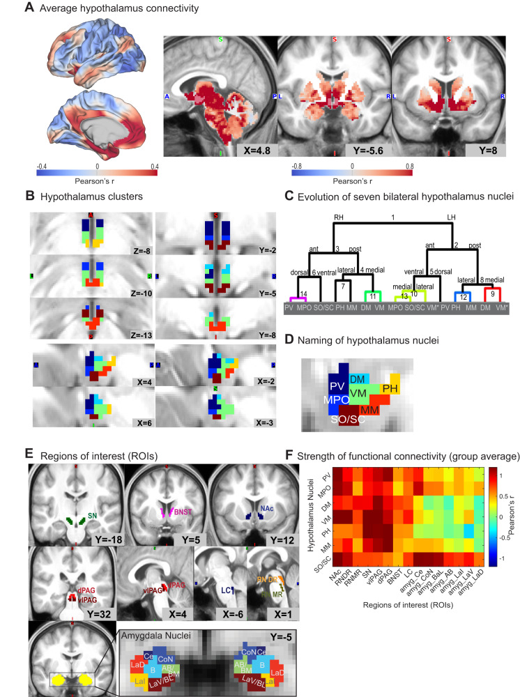
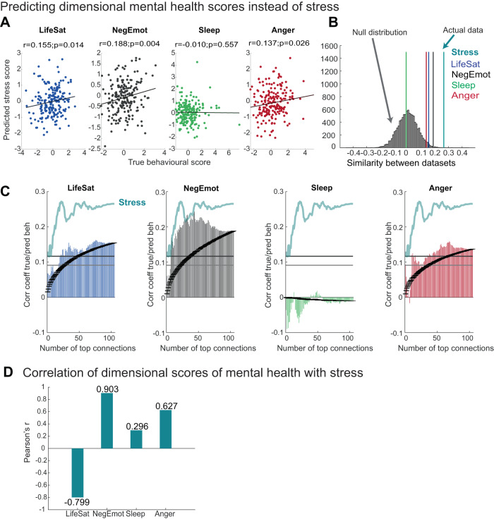
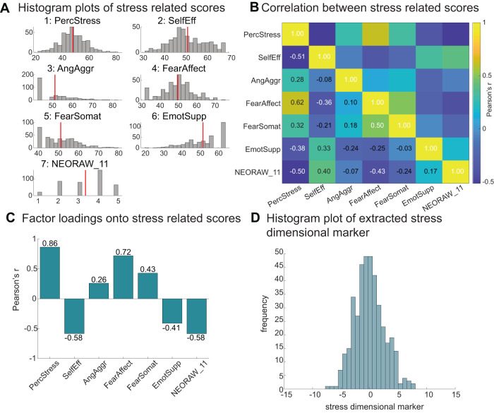
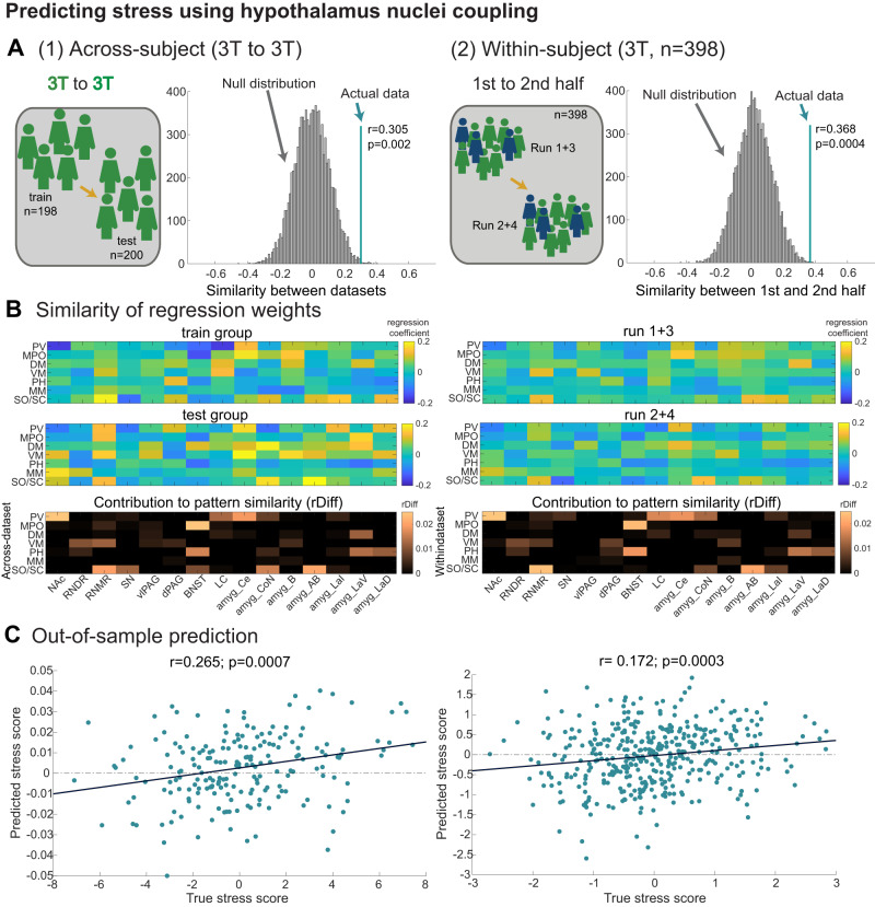
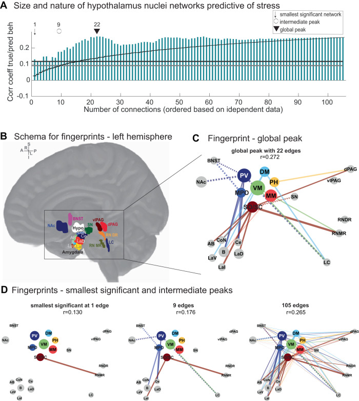
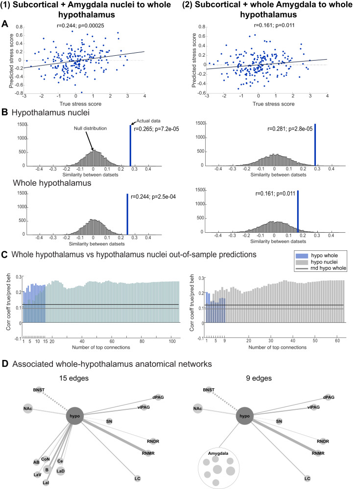
The hypothalamus is part of the hypothalamic-pituitary-adrenal axis which activates stress responses through release of cortisol. It is a small but heterogeneous structure comprising multiple nuclei. In vivo human neuroimaging has rarely succeeded in recording signals from individual hypothalamus nuclei. Here we use human resting-state fMRI (n = 498) with high spatial resolution to examine relationships between the functional connectivity of specific hypothalamic nuclei and a dimensional marker of prolonged stress. First, we demonstrate that we can parcellate the human hypothalamus into seven nuclei in vivo. Using the functional connectivity between these nuclei and other subcortical structures including the amygdala, we significantly predict stress scores out-of-sample. Predictions use 0.0015% of all possible brain edges, are specific to stress, and improve when using nucleus-specific compared to whole-hypothalamus connectivity. Thus, stress relates to connectivity changes in precise and functionally meaningful subcortical networks, which may be exploited in future studies using interventions in stress disorders.
下丘脑是下丘脑-垂体-肾上腺轴的一部分,通过释放皮质醇激活应激反应。它是一个小而异构的结构,包含多个核。在体内,人类神经影像学很少成功地从单个下丘脑核记录信号。在这里,我们使用具有高空间分辨率的人类静息状态 fMRI(n=498)来研究特定下丘脑核的功能连接与长期压力的维度标记之间的关系。首先,我们证明我们可以在体内将人类下丘脑分割成七个核。我们使用这些核与包括杏仁核在内的其他皮质下结构之间的功能连接,对压力评分进行了样本外预测。预测使用了所有可能脑边缘的 0.0015%,专门针对压力,并且当使用核特异性连接而不是整个下丘脑连接时会得到改善。因此,压力与精确和具有功能意义的皮质下网络中的连接变化有关,这可能在未来使用应激障碍的干预措施的研究中得到利用。