Wei Yingming, Wang Zhongxiu, Lei Lihong, Han Jiayin, Zhong Shuaiqi, Yang Xianyan, Gou Zhongru, Chen Lili
Department of Oral Medicine, The Second Affiliated Hospital, School of Medicine, Zhejiang University Hangzhou, 310008, China.
Bio-nanomaterials and Regenerative Medicine Research Division, Zhejiang-California International Nanosystem Institute, Zhejiang University, Hangzhou, 310058, China.
J Orthop Translat. 2024 Mar 13;45:88-99. doi: 10.1016/j.jot.2024.02.004. eCollection 2024 Mar.
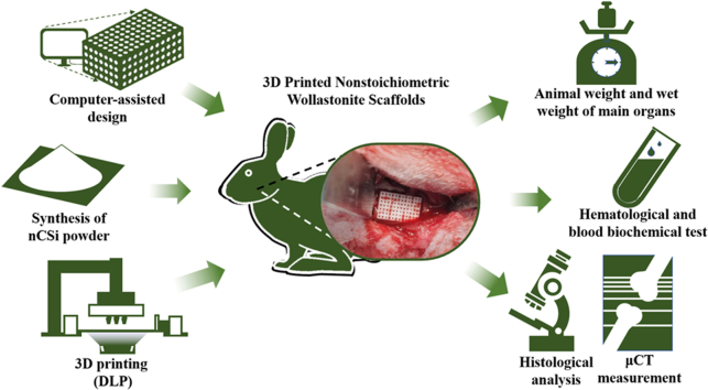
Alveolar bone destruction due to periodontal disease often requires a bone graft substitute to reconstruct the anatomical structures and biological functions of the bone tissue. Despite significant advances in the development of foreign ion-doped nonstoichiometric wollastonite bioceramics (CaSiO, nCSi) for alveolar bone regeneration over the past decade, the in vivo biosafety and osteogenesis of nCSi scaffolds remain uncertain. In this study, we developed a customized porous nCSi scaffold to investigate the in vivo biocompatibility and osteogenic properties of nCSi bioceramics.
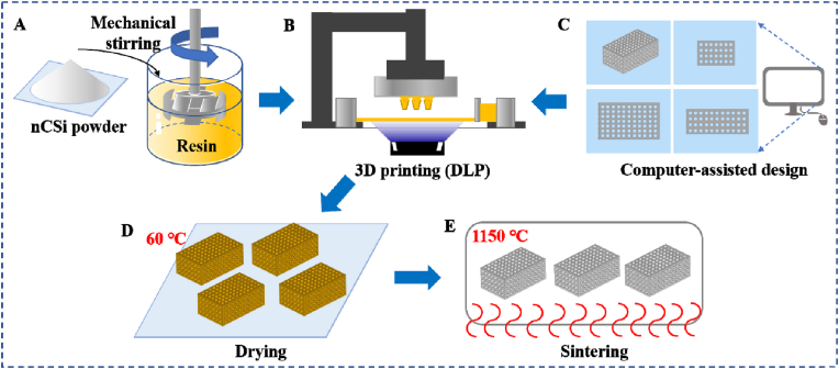
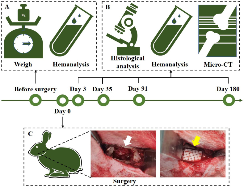
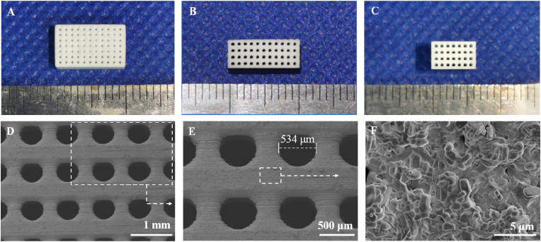
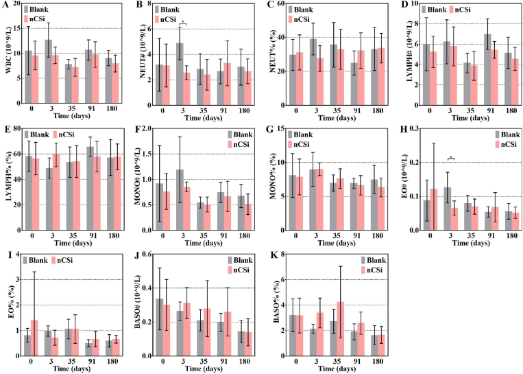
Six percent Mg-doped nCSi bioceramic scaffolds were fabricated by digital light processing (DLP), and the scaffold morphology, pore architecture, compressive strength, in vitro biodegradation, and apatite-forming ability of the bioceramic scaffolds were investigated systematically. Subsequently, an alveolar bone defect rabbit model was used to evaluate the biocompatibility and osteogenic efficacy of the nCSi bioceramics. Animal weight, hematological test, blood biochemical test, wet weight of the main organs, and pathological examination of the main organs were conducted. Micro-CT and histological staining were performed to analyze the osteogenic potential of the personalized bioceramic scaffolds.
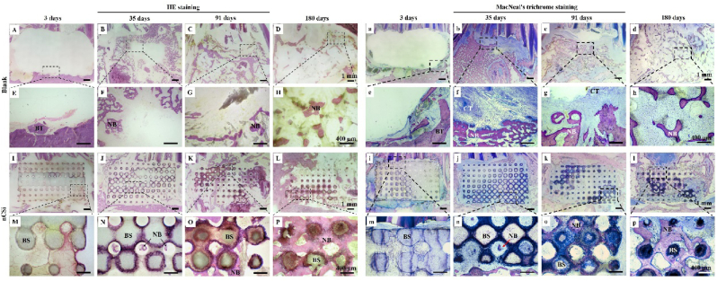
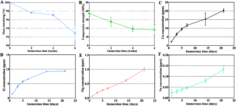
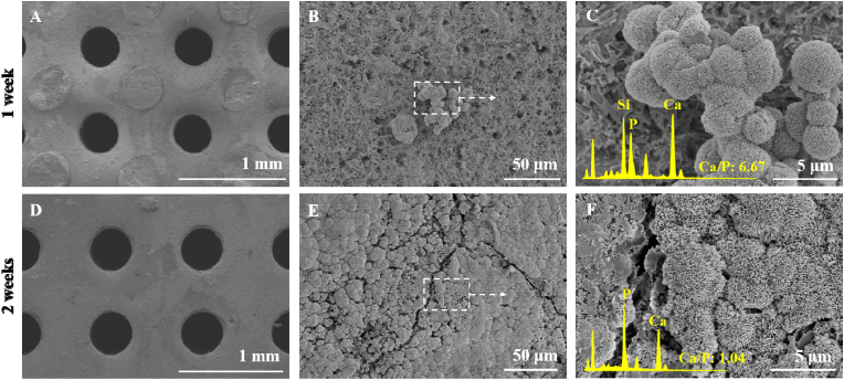
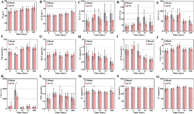
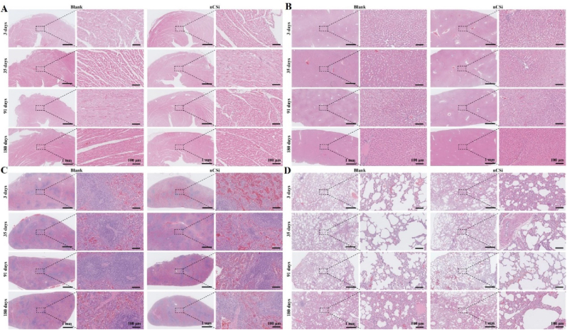
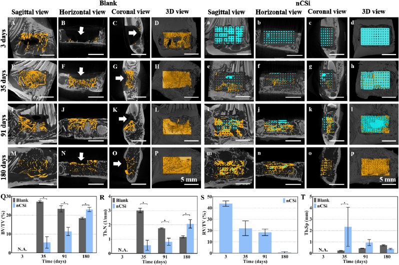
The nCSi scaffolds exhibited appreciable initial compressive strength (>30 MPa) and mild mechanical decay over time during in vitro biodissolution. In addition, the scaffolds induced apatite remineralization in SBF. Bioceramic scaffolds have been proven to have good biocompatibility in vivo after implantation into the alveolar bone defect of rabbits. No significant effects on the hematological indices, blood biochemical parameters, organ wet weight, or organ histopathology were detected from 3 to 180 days postoperatively. The porous scaffolds exhibited strong bone regeneration capability in the alveolar bone defect model of rabbits. Micro-CT and histological examination showed effective maintenance of bone morphology in the bioceramic scaffold group; however, depressed bone tissue was observed in the control group.
Our results suggest that personalized nCSi bioceramic scaffolds can be fabricated using the DLP technique. These newly developed strong bioceramic scaffolds exhibit good biocompatibility and osteogenic capability in vivo and have excellent potential as next-generation oral implants.
Tissue-engineered strategies for alveolar bone repair require a bone graft substitute with appreciable biocompatibility and osteogenic capability. This article provides a systematic investigation of the in vivo biosafety and osteogenic property of nCSi to further development of a silicate-based bioceramics materials for clinical applications.
牙周病导致的牙槽骨破坏通常需要骨替代物来重建骨组织的解剖结构和生物学功能。尽管在过去十年中,用于牙槽骨再生的外源性离子掺杂非化学计量硅灰石生物陶瓷(CaSiO₃,nCSi)的研发取得了显著进展,但nCSi支架在体内的生物安全性和成骨能力仍不确定。在本研究中,我们开发了一种定制的多孔nCSi支架,以研究nCSi生物陶瓷在体内的生物相容性和成骨特性。
采用数字光处理(DLP)技术制备了6%镁掺杂的nCSi生物陶瓷支架,并系统研究了支架的形态、孔隙结构、抗压强度、体外生物降解性和生物陶瓷支架的磷灰石形成能力。随后,使用牙槽骨缺损兔模型评估nCSi生物陶瓷的生物相容性和成骨效果。进行动物体重、血液学检测、血液生化检测、主要器官湿重和主要器官病理检查。采用Micro-CT和组织学染色分析个性化生物陶瓷支架的成骨潜力。
nCSi支架在体外生物溶解过程中表现出可观的初始抗压强度(>30MPa),且随着时间的推移机械性能轻度衰减。此外,该支架在模拟体液(SBF)中诱导了磷灰石再矿化。生物陶瓷支架植入兔牙槽骨缺损后,已被证明在体内具有良好的生物相容性。术后3至180天,未检测到对血液学指标、血液生化参数、器官湿重或器官组织病理学有显著影响。多孔支架在兔牙槽骨缺损模型中表现出强大的骨再生能力。Micro-CT和组织学检查显示生物陶瓷支架组的骨形态得到有效维持;然而,对照组观察到骨组织萎缩。
我们的结果表明,可以使用DLP技术制备个性化的nCSi生物陶瓷支架。这些新开发的高强度生物陶瓷支架在体内表现出良好的生物相容性和成骨能力,作为下一代口腔种植体具有巨大潜力。
牙槽骨修复的组织工程策略需要具有可观生物相容性和成骨能力的骨替代物。本文对nCSi的体内生物安全性和成骨特性进行了系统研究,以进一步开发用于临床应用的硅酸盐基生物陶瓷材料。