Department of Microbiology, University of Massachusetts, Amherst, MA, USA.
Division of Rheumatology, Immunity and Inflammation, Brigham and Women's Hospital, Harvard Medical School, Boston, MA, USA.
J Lipid Res. 2024 Jul;65(7):100533. doi: 10.1016/j.jlr.2024.100533. Epub 2024 Mar 24.
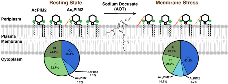
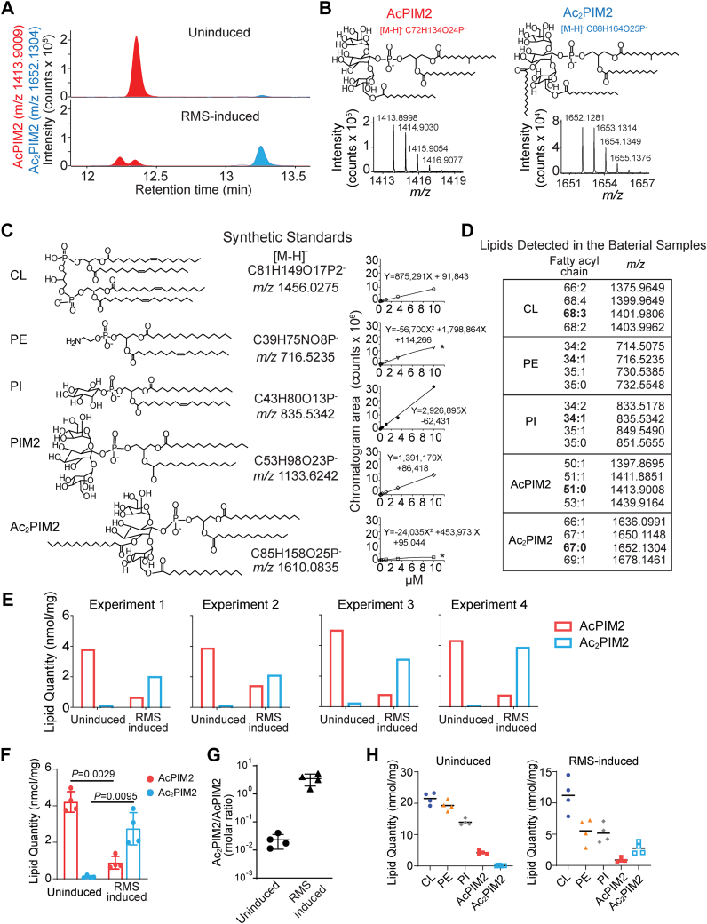
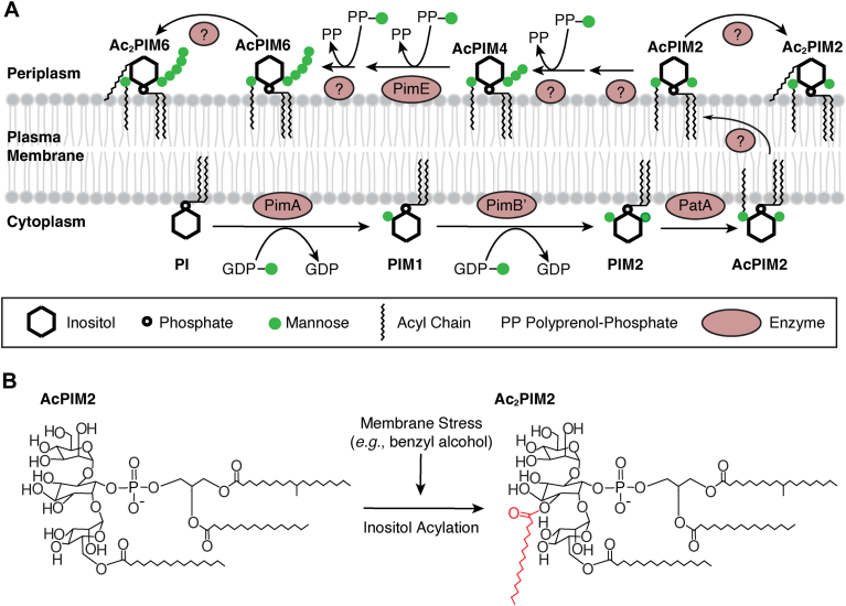
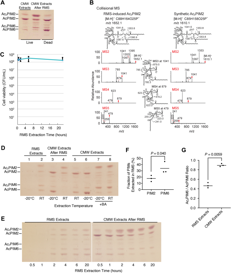
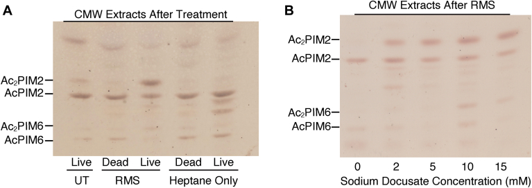
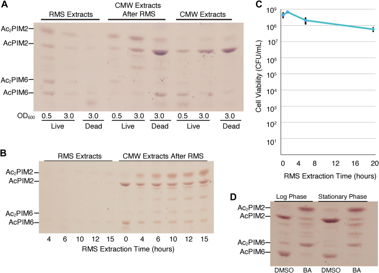
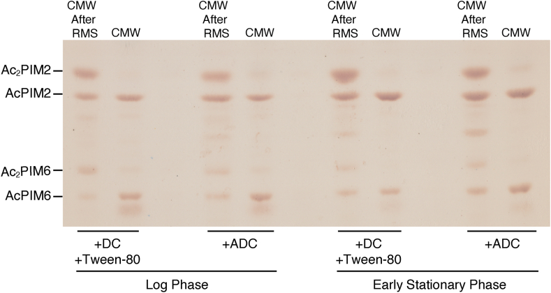
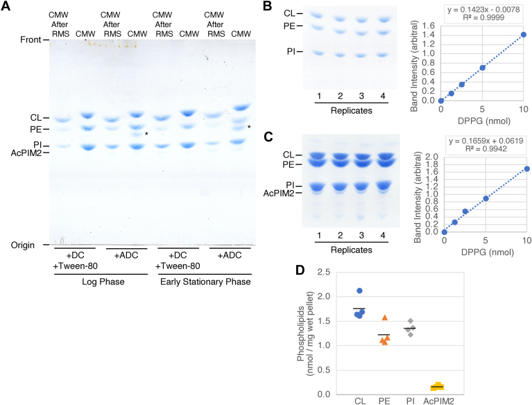
Mycobacterial plasma membrane, together with the peptidoglycan-arabinogalactan cell wall and waxy outer membrane, creates a robust permeability barrier against xenobiotics. The fact that several antituberculosis drugs target plasma membrane-embedded enzymes underscores the importance of the plasma membrane in bacterial physiology and pathogenesis. Nevertheless, its accurate phospholipid composition remains undefined, with conflicting reports on the abundance of phosphatidylinositol mannosides (PIMs), physiologically important glycolipids evolutionarily conserved among mycobacteria and related bacteria. Some studies indicate cardiolipin, phosphatidylethanolamine, and phosphatidylinositol as dominant structural phospholipids. Conversely, some suggest PIMs dominate the plasma membrane. A striking example of the latter is the use of reverse micelle extraction, showing diacyl phosphatidylinositol dimannoside (AcPIM2) as the most abundant phospholipid in a model organism, Mycobacterium smegmatis. Our recent work reveals a rapid response mechanism to membrane-fluidizing stress in mycobacterial plasma membrane: monoacyl phosphatidylinositol dimannoside and hexamannoside (AcPIM2 and AcPIM6) are converted to diacyl forms (AcPIM2 and AcPIM6). Given the dynamic nature of PIMs, we aimed to resolve the conflicting data in the literature. We show that unstressed M. smegmatis lacks an AcPIM2-dominated plasma membrane. AcPIM2 accumulation is induced by experimental conditions involving sodium docusate, a component of the reverse micellar solution. Using chemically synthesized PIMs as standards, we accurately quantified phospholipid ratio in M. smegmatis through liquid chromatography-mass spectrometry, revealing that mycobacterial plasma membrane is dominated by cardiolipin, phosphatidylethanolamine, and phosphatidylinositol. PIMs are quantitatively minor but responsive to environmental stresses in M. smegmatis. Our study paves the way for accurate modeling of mycobacterial plasma membrane.
分枝杆菌的质膜与肽聚糖-阿拉伯半乳聚糖细胞壁和蜡质外膜一起,为外来物质创造了一个强大的渗透屏障。事实上,几种抗结核药物的靶点是质膜嵌入的酶,这突显了质膜在细菌生理学和发病机制中的重要性。然而,其准确的磷脂组成仍未确定,关于磷脂酰肌醇甘露糖苷(PIMs)的丰度存在相互矛盾的报告,PIMs 是生理上重要的糖脂,在分枝杆菌和相关细菌中进化上保守。一些研究表明心磷脂、磷脂酰乙醇胺和磷脂酰肌醇是主要的结构磷脂。相反,一些研究表明 PIMs 主导质膜。后者的一个突出例子是使用反胶束提取,表明二酰基磷脂酰肌醇二甘露糖苷(AcPIM2)是模式生物分枝杆菌中的最丰富磷脂。我们最近的工作揭示了分枝杆菌质膜对膜流动性胁迫的快速反应机制:单酰基磷脂酰肌醇二甘露糖苷和六甘露糖苷(AcPIM2 和 AcPIM6)转化为二酰基形式(AcPIM2 和 AcPIM6)。鉴于 PIMs 的动态性质,我们旨在解决文献中的矛盾数据。我们表明,未受应激的耻垢分枝杆菌缺乏以 AcPIM2 为主导的质膜。AcPIM2 的积累是由涉及十二烷基硫酸钠的实验条件诱导的,十二烷基硫酸钠是反胶束溶液的一种成分。我们使用化学合成的 PIMs 作为标准,通过液相色谱-质谱法准确地定量了耻垢分枝杆菌中的磷脂比例,结果表明分枝杆菌的质膜主要由心磷脂、磷脂酰乙醇胺和磷脂酰肌醇组成。PIMs 的数量较少,但对分枝杆菌中的环境应激有反应。我们的研究为准确建模分枝杆菌质膜铺平了道路。