• 文献检索
  • 文档翻译
  • 深度研究
  • 学术资讯
  • Suppr Zotero 插件Zotero 插件
  • 邀请有礼
  • 套餐&价格
  • 历史记录
应用&插件
Suppr Zotero 插件Zotero 插件浏览器插件Mac 客户端Windows 客户端微信小程序
定价
高级版会员购买积分包购买API积分包
服务
文献检索文档翻译深度研究API 文档MCP 服务
关于我们
关于 Suppr公司介绍联系我们用户协议隐私条款
关注我们

Suppr 超能文献

核心技术专利:CN118964589B侵权必究
粤ICP备2023148730 号-1Suppr @ 2026

文献检索

告别复杂PubMed语法,用中文像聊天一样搜索,搜遍4000万医学文献。AI智能推荐,让科研检索更轻松。

立即免费搜索

文件翻译

保留排版,准确专业,支持PDF/Word/PPT等文件格式,支持 12+语言互译。

免费翻译文档

深度研究

AI帮你快速写综述,25分钟生成高质量综述,智能提取关键信息,辅助科研写作。

立即免费体验

颅内动脉瘤血管内治疗后的影像学随访策略:文献综述与指南建议

Imaging follow-up strategy after endovascular treatment of Intracranial aneurysms: A literature review and guideline recommendations.

作者信息

Cho Yong-Hwan, Choi Jaehyung, Huh Chae-Wook, Kim Chang Hyeun, Chang Chul Hoon, Kwon Soon Chan, Kim Young Woo, Sheen Seung Hun, Park Sukh Que, Ko Jun Kyeung, Ha Sung-Kon, Jeong Hae Woong, Kang Hyen Seung

机构信息

Department of Neurosurgery, Dong-A University Hospital, Busan, Korea.

Department of Neurosurgery, Busan-Ulsan Regional Cardiocerebrovascular Center, Dong-A University Hospital, Busan, Korea.

出版信息

J Cerebrovasc Endovasc Neurosurg. 2024 Mar;26(1):1-10. doi: 10.7461/jcen.2024.E2023.08.008. Epub 2024 Mar 25.

DOI:10.7461/jcen.2024.E2023.08.008
PMID:38523549
原文链接:https://pmc.ncbi.nlm.nih.gov/articles/PMC10995472/
Abstract

OBJECTIVE

Endovascular coil embolization is the primary treatment modality for intracranial aneurysms. However, its long-term durability remains of concern, with a considerable proportion of cases requiring aneurysm reopening and retreatment. Therefore, establishing optimal follow-up imaging protocols is necessary to ensure a durable occlusion. This study aimed to develop guidelines for follow-up imaging strategies after endovascular treatment of intracranial aneurysms.

METHODS

A committee comprising members of the Korean Neuroendovascular Society and other relevant societies was formed. A literature review and analyses of the major published guidelines were conducted to gather evidence. A panel of 40 experts convened to achieve a consensus on the recommendations using the modified Delphi method.

RESULTS

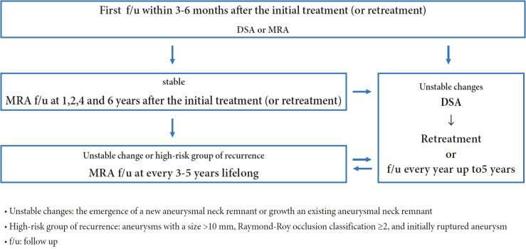
The panel members reached the following consensus: 1. Schedule the initial follow-up imaging within 3-6 months of treatment. 2. Noninvasive imaging modalities, such as three-dimensional time-of-flight magnetic resonance angiography (MRA) or contrast-enhanced MRA, are alternatives to digital subtraction angiography (DSA) during the first follow-up. 3. Schedule mid-term follow-up imaging at 1, 2, 4, and 6 years after the initial treatment. 4. If noninvasive imaging reveals unstable changes in the treated aneurysms, DSA should be considered. 5. Consider late-term follow-up imaging every 3-5 years for lifelong monitoring of patients with unstable changes or at high risk of recurrence.

CONCLUSIONS

The guidelines aim to provide physicians with the information to make informed decisions and provide patients with high-quality care. However, owing to a lack of specific recommendations and scientific data, these guidelines are based on expert consensus and should be considered in conjunction with individual patient characteristics and circumstances.

摘要

目的

血管内弹簧圈栓塞术是颅内动脉瘤的主要治疗方式。然而,其长期耐久性仍令人担忧,相当一部分病例需要再次打开动脉瘤并进行再次治疗。因此,制定最佳的随访成像方案对于确保持久闭塞是必要的。本研究旨在制定颅内动脉瘤血管内治疗后随访成像策略的指南。

方法

成立了一个由韩国神经血管内介入学会成员和其他相关学会成员组成的委员会。进行了文献综述并分析了主要已发表的指南以收集证据。召集了40位专家组成的小组,使用改良的德尔菲法就这些建议达成共识。

结果

小组成员达成了以下共识:1. 在治疗后3 - 6个月安排首次随访成像。2. 在首次随访期间,非侵入性成像方式,如三维时间飞跃磁共振血管造影(MRA)或对比增强MRA,可作为数字减影血管造影(DSA)的替代方法。3. 在初始治疗后的1、2、4和6年安排中期随访成像。4. 如果非侵入性成像显示治疗的动脉瘤有不稳定变化,应考虑进行DSA检查。5. 对于有不稳定变化或复发高风险的患者,考虑每3 - 5年进行晚期随访成像,以进行终身监测。

结论

这些指南旨在为医生提供信息,以便做出明智的决策,并为患者提供高质量的护理。然而,由于缺乏具体建议和科学数据依据,这些指南基于专家共识,应结合个体患者的特征和情况加以考虑。

https://cdn.ncbi.nlm.nih.gov/pmc/blobs/a66c/10995472/724e9707bc03/jcen-2024-e2023-08-008f1.jpg

相似文献

1
Imaging follow-up strategy after endovascular treatment of Intracranial aneurysms: A literature review and guideline recommendations.颅内动脉瘤血管内治疗后的影像学随访策略:文献综述与指南建议
J Cerebrovasc Endovasc Neurosurg. 2024 Mar;26(1):1-10. doi: 10.7461/jcen.2024.E2023.08.008. Epub 2024 Mar 25.
2
The clinical value of ceMRA versus DSA for follow-up of intracranial aneurysms treated by coil embolization: an assessment of occlusion classifications and impact on treatment decisions.ceMRA 与 DSA 对颅内动脉瘤线圈栓塞治疗后随访的临床价值:闭塞分类评估及其对治疗决策的影响。
Eur Radiol. 2021 Jun;31(6):4104-4113. doi: 10.1007/s00330-020-07492-3. Epub 2020 Nov 21.
3
Magnetic resonance angiography or digital subtraction catheter angiography for follow-up of coiled aneurysms: do we need both?磁共振血管造影或数字减影导管血管造影用于盘绕动脉瘤的随访:两者都需要吗?
J Med Imaging Radiat Oncol. 2015 Apr;59(2):163-9. doi: 10.1111/1754-9485.12288.
4
Time-of-flight MR angiography at 3T versus digital subtraction angiography in the imaging follow-up of 51 intracranial aneurysms treated with coils.3T 时飞越磁共振血管造影与数字减影血管造影在 51 例颅内动脉瘤弹簧圈治疗后影像学随访中的对比研究。
Eur J Radiol. 2009 Dec;72(3):365-9. doi: 10.1016/j.ejrad.2008.08.005. Epub 2008 Sep 21.
5
Follow-up after embolization of ruptured intracranial aneurysms: a prospective comparison of two-dimensional digital subtraction angiography, three-dimensional digital subtraction angiography, and time-of-flight magnetic resonance angiography.破裂颅内动脉瘤栓塞后随访:二维数字减影血管造影、三维数字减影血管造影和时间飞跃磁共振血管造影的前瞻性比较。
Neuroradiology. 2012 Nov;54(11):1253-60. doi: 10.1007/s00234-012-1030-z. Epub 2012 Apr 10.
6
MRA versus DSA for the follow-up imaging of intracranial aneurysms treated using endovascular techniques: a meta-analysis.MRA 与 DSA 用于血管内治疗颅内动脉瘤后的随访成像:一项荟萃分析。
J Neurointerv Surg. 2019 Oct;11(10):1009-1014. doi: 10.1136/neurintsurg-2019-014936. Epub 2019 May 2.
7
Feasibility of magnetic resonance angiography (MRA) follow-up as the primary imaging modality after coiling of intracranial aneurysms.颅内动脉瘤栓塞术后磁共振血管造影(MRA)随访作为主要成像方式的可行性。
Acta Radiol. 2010 Mar;51(2):226-32. doi: 10.3109/02841850903436642.
8
MR angiography at 3T versus digital subtraction angiography in the follow-up of intracranial aneurysms treated with detachable coils.3T磁共振血管造影与数字减影血管造影在可脱性弹簧圈治疗颅内动脉瘤随访中的比较
AJNR Am J Neuroradiol. 2005 Jun-Jul;26(6):1349-56.
9
No advantage of time-of-flight magnetic resonance angiography at 3 Tesla compared to 1.5 Tesla in the follow-up after endovascular treatment of cerebral aneurysms.在脑动脉瘤血管内治疗后的随访中,3特斯拉飞行时间磁共振血管造影与1.5特斯拉相比并无优势。
Neuroradiology. 2008 Oct;50(10):855-61. doi: 10.1007/s00234-008-0413-7. Epub 2008 Jun 4.
10
Enhanced vessel wall magnetic resonance imaging in the follow-up of intracranial aneurysms treated with flow diversion.血流导向治疗颅内动脉瘤随访中的增强血管壁磁共振成像
Eur Radiol. 2024 Feb;34(2):833-841. doi: 10.1007/s00330-023-10094-4. Epub 2023 Aug 15.

引用本文的文献

1
Gap between the clip and coil mass as a predictor of aneurysm recurrence and occlusion: illustrative case.夹闭器与弹簧圈团块之间的间隙作为动脉瘤复发和闭塞的预测指标:病例说明
J Neurosurg Case Lessons. 2025 Aug 4;10(5). doi: 10.3171/CASE25286.
2
Complex Anatomy, Advanced Techniques: Microsurgical Clipping of a Ruptured Hypophyseal Artery Aneurysm.复杂解剖结构与先进技术:破裂垂体动脉动脉瘤的显微外科夹闭术
J Clin Med. 2025 Mar 29;14(7):2361. doi: 10.3390/jcm14072361.
3
Intracranial Aneurysms Treated with a Novel Coated Low-Profile Flow Diverter (p48 HPC)-A Single-Center Experience and an Illustrative Case Series.

本文引用的文献

1
The sensitivity and specificity of TOF-MRA compared with DSA in the follow-up of treated intracranial aneurysms.TOF-MRA 与 DSA 在治疗后颅内动脉瘤随访中的敏感性和特异性比较。
J Neurointerv Surg. 2021 Dec;13(12):1172-1179. doi: 10.1136/neurintsurg-2020-016788. Epub 2021 Feb 25.
2
MRA versus DSA for the follow-up imaging of intracranial aneurysms treated using endovascular techniques: a meta-analysis.MRA 与 DSA 用于血管内治疗颅内动脉瘤后的随访成像:一项荟萃分析。
J Neurointerv Surg. 2019 Oct;11(10):1009-1014. doi: 10.1136/neurintsurg-2019-014936. Epub 2019 May 2.
3
Pointwise Encoding Time Reduction with Radial Acquisition with Subtraction-Based MRA during the Follow-Up of Stent-Assisted Coil Embolization of Anterior Circulation Aneurysms.
使用新型涂层低轮廓血流导向装置(p48 HPC)治疗颅内动脉瘤——单中心经验及病例系列展示
Brain Sci. 2025 Jan 3;15(1):42. doi: 10.3390/brainsci15010042.
4
Clipping aneurysms via a fully endoscopic transcranial approach.通过全内镜经颅入路夹闭动脉瘤。
Sci Rep. 2024 Dec 30;14(1):32134. doi: 10.1038/s41598-024-83958-4.
支架辅助线圈栓塞前循环动脉瘤随访中基于减法的 MRA 的径向采集点编码时间缩短。
AJNR Am J Neuroradiol. 2019 May;40(5):815-819. doi: 10.3174/ajnr.A6035. Epub 2019 Apr 11.
4
Usefulness of contrast-enhanced and TOF MR angiography for follow-up after low-profile stent-assisted coil embolization of intracranial aneurysms.对比增强磁共振血管造影和时间飞跃法磁共振血管造影在颅内动脉瘤低轮廓支架辅助弹簧圈栓塞术后随访中的应用价值。
Interv Neuroradiol. 2018 Dec;24(6):655-661. doi: 10.1177/1591019918785910. Epub 2018 Jul 5.
5
Imaging Follow-Up of Intracranial Aneurysms Treated by Endovascular Means: Why, When, and How?血管内治疗颅内动脉瘤的影像随访:为何、何时以及如何进行?
Stroke. 2016 May;47(5):1407-12. doi: 10.1161/STROKEAHA.115.011414. Epub 2016 Mar 29.
6
The Barrow Ruptured Aneurysm Trial: 6-year results.巴罗破裂动脉瘤试验:6年结果。
J Neurosurg. 2015 Sep;123(3):609-17. doi: 10.3171/2014.9.JNS141749. Epub 2015 Jun 26.
7
Intracranial Aneurysms: Recurrences More than 10 Years after Endovascular Treatment-A Prospective Cohort Study, Systematic Review, and Meta-Analysis.颅内动脉瘤:血管内治疗 10 年以上后的复发——一项前瞻性队列研究、系统评价和荟萃分析。
Radiology. 2015 Oct;277(1):173-80. doi: 10.1148/radiol.2015142496. Epub 2015 Jun 9.
8
Assessing Blood Flow in an Intracranial Stent: A Feasibility Study of MR Angiography Using a Silent Scan after Stent-Assisted Coil Embolization for Anterior Circulation Aneurysms.评估颅内支架内的血流:一项关于支架辅助弹簧圈栓塞治疗前循环动脉瘤后使用静音扫描进行磁共振血管造影的可行性研究。
AJNR Am J Neuroradiol. 2015 May;36(5):967-70. doi: 10.3174/ajnr.A4199. Epub 2014 Dec 18.
9
The durability of endovascular coiling versus neurosurgical clipping of ruptured cerebral aneurysms: 18 year follow-up of the UK cohort of the International Subarachnoid Aneurysm Trial (ISAT).破裂性脑动脉瘤血管内栓塞与神经外科夹闭术的耐久性:国际蛛网膜下腔动脉瘤试验(ISAT)英国队列的18年随访
Lancet. 2015 Feb 21;385(9969):691-7. doi: 10.1016/S0140-6736(14)60975-2. Epub 2014 Oct 28.
10
Follow-up of intracranial aneurysms treated by flow diverter: comparison of three-dimensional time-of-flight MR angiography (3D-TOF-MRA) and contrast-enhanced MR angiography (CE-MRA) sequences with digital subtraction angiography as the gold standard.血流导向装置治疗颅内动脉瘤的随访:以数字减影血管造影为金标准比较三维时间飞跃磁共振血管造影(3D-TOF-MRA)和对比增强磁共振血管造影(CE-MRA)序列
J Neurointerv Surg. 2016 Jan;8(1):81-6. doi: 10.1136/neurintsurg-2014-011449. Epub 2014 Oct 28.