• 文献检索
  • 文档翻译
  • 深度研究
  • 学术资讯
  • Suppr Zotero 插件Zotero 插件
  • 邀请有礼
  • 套餐&价格
  • 历史记录
应用&插件
Suppr Zotero 插件Zotero 插件浏览器插件Mac 客户端Windows 客户端微信小程序
定价
高级版会员购买积分包购买API积分包
服务
文献检索文档翻译深度研究API 文档MCP 服务
关于我们
关于 Suppr公司介绍联系我们用户协议隐私条款
关注我们

Suppr 超能文献

核心技术专利:CN118964589B侵权必究
粤ICP备2023148730 号-1Suppr @ 2026

文献检索

告别复杂PubMed语法,用中文像聊天一样搜索,搜遍4000万医学文献。AI智能推荐,让科研检索更轻松。

立即免费搜索

文件翻译

保留排版,准确专业,支持PDF/Word/PPT等文件格式,支持 12+语言互译。

免费翻译文档

深度研究

AI帮你快速写综述,25分钟生成高质量综述,智能提取关键信息,辅助科研写作。

立即免费体验

用于跨越踝关节外固定的第二根跟骨针的添加

Addition of a Second Calcaneal Pin for Spanning Ankle External Fixation.

作者信息

Deluca Meridith, Hoffman Brett A, Serdahely Kevin, Ravi Sreeram, Sanford Christopher

机构信息

Orthopedics, The University of Toledo College of Medicine and Life Sciences, Toledo, USA.

Orthopedics, Penn State Health Milton S. Hershey Medical Center, State College, USA.

出版信息

Cureus. 2024 Mar 1;16(3):e55312. doi: 10.7759/cureus.55312. eCollection 2024 Mar.

DOI:10.7759/cureus.55312
PMID:38559505
原文链接:https://pmc.ncbi.nlm.nih.gov/articles/PMC10981796/
Abstract

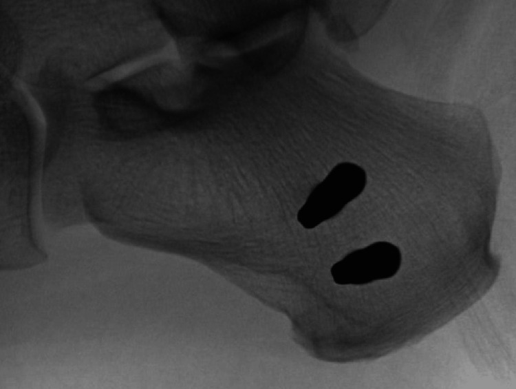
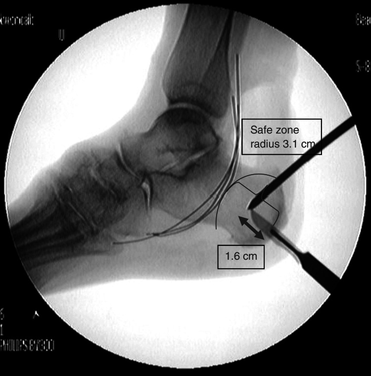
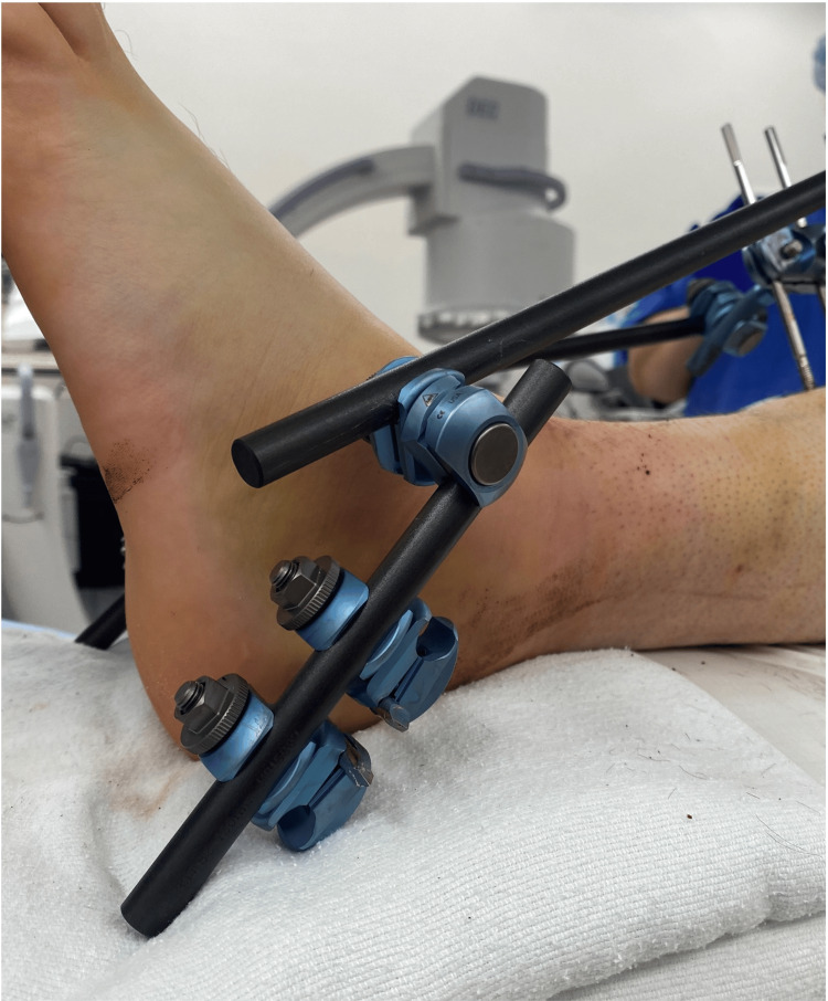
Spanning ankle external fixation is a commonly used technique for the treatment of fractures of the lower extremity. Traditionally, a single pin is placed in the safe zone of the calcaneus to provide a point of traction for fracture reduction and stabilization. Complications include infection and pin loosening with subsequent loss of fracture reduction. We aim to highlight the benefits and techniques of adding a second calcaneal pin to reduce the likelihood of infection, pin loosening, and possible loss of fracture reduction. Using the standard medial-to-lateral placement technique, two centrally threaded Schanz pins were placed within the safe zone of the calcaneus approximately 2 cm apart and were connected by clamps and a short carbon fiber rod. The remainder of the external fixation apparatus is assembled using a standard technique after obtaining fracture reduction. There is an increased incidence of infection and pin loosening with decreased bone quality and a longer duration within an external fixator. The addition of a second calcaneal pin can be used to reduce the incidence of pin loosening and associated sequela, especially in patients with decreased bone quality, thus improving outcomes for patients undergoing spanning ankle external fixation.

摘要

跨踝关节外固定是治疗下肢骨折常用的技术。传统上,在跟骨安全区置入一枚钢针,为骨折复位和固定提供牵引点。并发症包括感染和钢针松动,继而导致骨折复位丢失。我们旨在强调增加一枚跟骨钢针的益处和技术,以降低感染、钢针松动以及可能的骨折复位丢失的可能性。采用标准的从内侧到外侧的置入技术,在跟骨安全区内相距约2厘米处置入两枚中心螺纹斯氏针,并用夹具和短碳纤维棒连接。在获得骨折复位后,使用标准技术组装外固定装置的其余部分。随着骨质下降和外固定器使用时间延长,感染和钢针松动的发生率会增加。增加一枚跟骨钢针可用于降低钢针松动及相关后遗症的发生率,尤其是在骨质下降的患者中,从而改善接受跨踝关节外固定患者的治疗效果。

https://cdn.ncbi.nlm.nih.gov/pmc/blobs/0236/10981796/88f27bc23efb/cureus-0016-00000055312-i03.jpg
https://cdn.ncbi.nlm.nih.gov/pmc/blobs/0236/10981796/e63fc5341af9/cureus-0016-00000055312-i01.jpg
https://cdn.ncbi.nlm.nih.gov/pmc/blobs/0236/10981796/1568363428fa/cureus-0016-00000055312-i02.jpg
https://cdn.ncbi.nlm.nih.gov/pmc/blobs/0236/10981796/88f27bc23efb/cureus-0016-00000055312-i03.jpg
https://cdn.ncbi.nlm.nih.gov/pmc/blobs/0236/10981796/e63fc5341af9/cureus-0016-00000055312-i01.jpg
https://cdn.ncbi.nlm.nih.gov/pmc/blobs/0236/10981796/1568363428fa/cureus-0016-00000055312-i02.jpg
https://cdn.ncbi.nlm.nih.gov/pmc/blobs/0236/10981796/88f27bc23efb/cureus-0016-00000055312-i03.jpg

相似文献

1
Addition of a Second Calcaneal Pin for Spanning Ankle External Fixation.用于跨越踝关节外固定的第二根跟骨针的添加
Cureus. 2024 Mar 1;16(3):e55312. doi: 10.7759/cureus.55312. eCollection 2024 Mar.
2
Monoaxial external fixation of the calcaneus: An anatomical study assessing the safety of monoaxial pin insertion.跟骨单轴外固定:一项评估单轴钢针插入安全性的解剖学研究。
Injury. 2016 Oct;47(10):2091-2096. doi: 10.1016/j.injury.2016.06.024. Epub 2016 Jun 23.
3
Calcaneus traction compression with orthopaedic reduction forceps combined with percutaneous minimally invasive treatment of intra-articular calcaneal fractures: An analysis of efficacy.跟骨牵引压缩配合骨科复位钳联合经皮微创治疗关节内跟骨骨折:疗效分析。
Biomed Pharmacother. 2020 Aug;128:110295. doi: 10.1016/j.biopha.2020.110295. Epub 2020 Jun 15.
4
Could percutaneous fixation with crossed Schanz pins be an alternative to open reduction in the treatment of intra-articular calcaneal fractures?经皮交叉斯氏针固定是否可作为治疗关节内跟骨骨折的切开复位的替代方法?
Int Orthop. 2021 Mar;45(3):731-741. doi: 10.1007/s00264-021-04944-3. Epub 2021 Jan 30.
5
The safety of forefoot metatarsal pins in external fixation of the lower extremity.下肢外固定中前足跖骨钢针的安全性
J Bone Joint Surg Am. 2008 Mar;90(3):560-4. doi: 10.2106/JBJS.G.00743.
6
External fixation of the calcaneus and talus: an anatomical study for safe pin insertion.跟骨和距骨的外固定:安全进针的解剖学研究
J Orthop Trauma. 1996;10(7):487-91. doi: 10.1097/00005131-199610000-00007.
7
[Case-control study on calcaneal traction and external fixator fixation of fractional delayed surgery for type C Pilon Fracture].[跟骨牵引联合外固定架固定对C型Pilon骨折延期手术疗效的病例对照研究]
Zhongguo Gu Shang. 2020 Mar 25;33(3):203-8. doi: 10.12200/j.issn.1003-0034.2020.03.003.
8
External Fixation Principles and Overview外固定原理与概述
9
Percutaneous pin placement in the medial calcaneus: is anywhere safe?经皮跟骨内侧穿针:任何部位都安全吗?
J Orthop Trauma. 2004 Sep;18(8 Suppl):S39-42. doi: 10.1097/00005131-200409001-00006.
10
Percutaneous pin placement in the medial calcaneus: is anywhere safe?经皮跟骨内侧克氏针置入:任何部位都安全吗?
J Orthop Trauma. 2002 Jan;16(1):26-9. doi: 10.1097/00005131-200201000-00006.

本文引用的文献

1
Reduction of pin tract infections during external fixation using cadexomer iodine.使用碘卡多曲减少外固定期间的针道感染。
J Exp Orthop. 2020 Nov 7;7(1):88. doi: 10.1186/s40634-020-00305-y.
2
Surgical Treatment and Outcomes of Calcaneal Osteomyelitis in Adults: A Systematic Review.成人跟骨骨髓炎的外科治疗及结果:一项系统评价
J Bone Jt Infect. 2019 May 21;4(3):146-154. doi: 10.7150/jbji.34452. eCollection 2019.
3
Calcaneal tuberosity fractures through prior Schantz pin sites in patients with diabetic neuropathy.糖尿病性神经病变患者经既往斯坎茨针穿刺部位的跟骨结节骨折
Foot (Edinb). 2019 Jun;39:96-99. doi: 10.1016/j.foot.2019.02.011. Epub 2019 Mar 2.
4
Hydroxyapatite pins for external fixation: is there sufficient evidence to prove that coated pins are less likely to be replaced prematurely?用于外固定的羟基磷灰石针:是否有足够证据证明涂层针过早更换的可能性较小?
Acta Orthop Traumatol Turc. 2015;49(4):410-5. doi: 10.3944/AOTT.2015.14.0206.
5
Bisphosphonate-coated external fixation pins appear similar to hydroxyapatite-coated pins in the tibial metaphysis and to uncoated pins in the shaft.双膦酸盐涂层外固定钉在胫骨干骺端与羟基磷灰石涂层钉相似,而在骨干处与未涂层钉相似。
Acta Orthop. 2013 Jun;84(3):314-8. doi: 10.3109/17453674.2013.797315. Epub 2013 Apr 28.
6
Tibial shaft fractures - management and treatment options. A review of the current literature.胫骨干骨折——管理与治疗选择。当前文献综述。
Acta Chir Orthop Traumatol Cech. 2012;79(6):499-505.
7
Bone properties affect loosening of half-pin external fixators at the pin-bone interface.骨骼特性会影响半针外固定器在针骨界面处的松动。
Injury. 2012 Oct;43(10):1764-70. doi: 10.1016/j.injury.2012.07.001. Epub 2012 Jul 28.
8
Calcaneal traction pin placement simplified: a cadaveric study.跟骨牵引针置钉简化:尸体研究。
Foot Ankle Int. 2011 Jun;32(6):651-5. doi: 10.3113/FAI.2011.0651.
9
The "safe zone" in medial percutaneous calcaneal pin placement.经皮跟骨内侧克氏针置入的“安全区”
Clin Anat. 2009 May;22(4):523-9. doi: 10.1002/ca.20778.
10
The safety of forefoot metatarsal pins in external fixation of the lower extremity.下肢外固定中前足跖骨钢针的安全性
J Bone Joint Surg Am. 2008 Mar;90(3):560-4. doi: 10.2106/JBJS.G.00743.