Department of Neurology, Massachusetts General Hospital, Boston, MA, 02114, USA.
MassGeneral Institute for Neurodegenerative Disease, 114 16th St., Charlestown, MA, 02129, USA.
Acta Neuropathol. 2024 Apr 3;147(1):66. doi: 10.1007/s00401-024-02712-2.
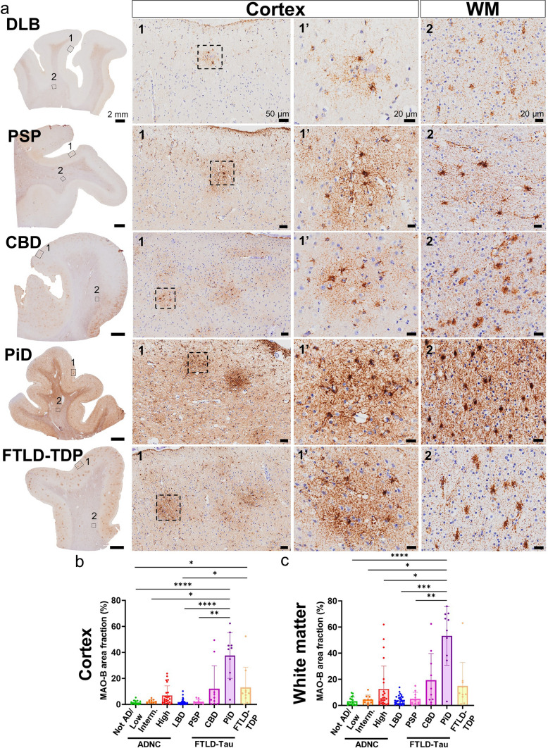
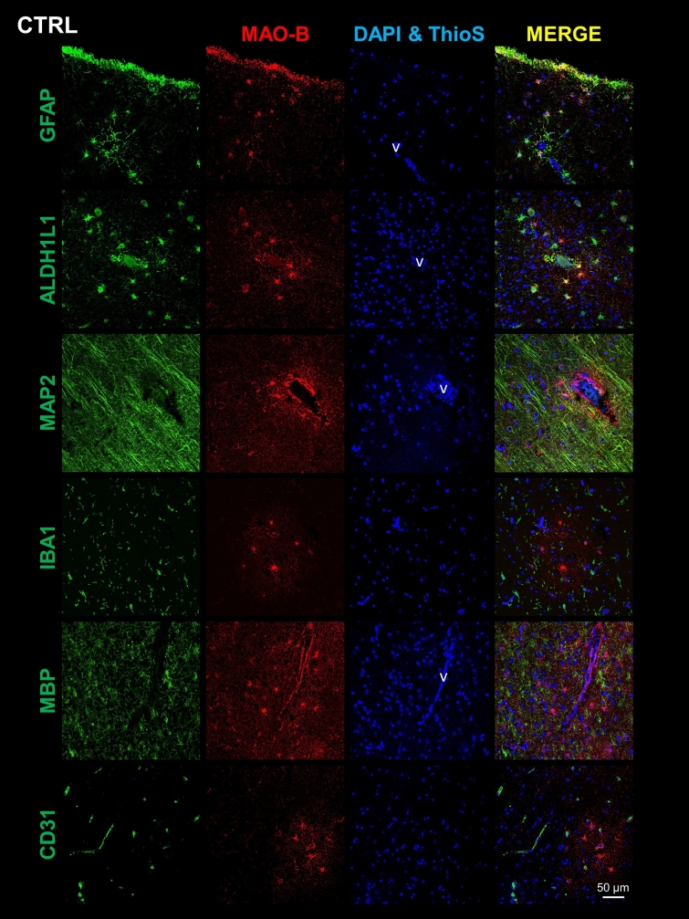
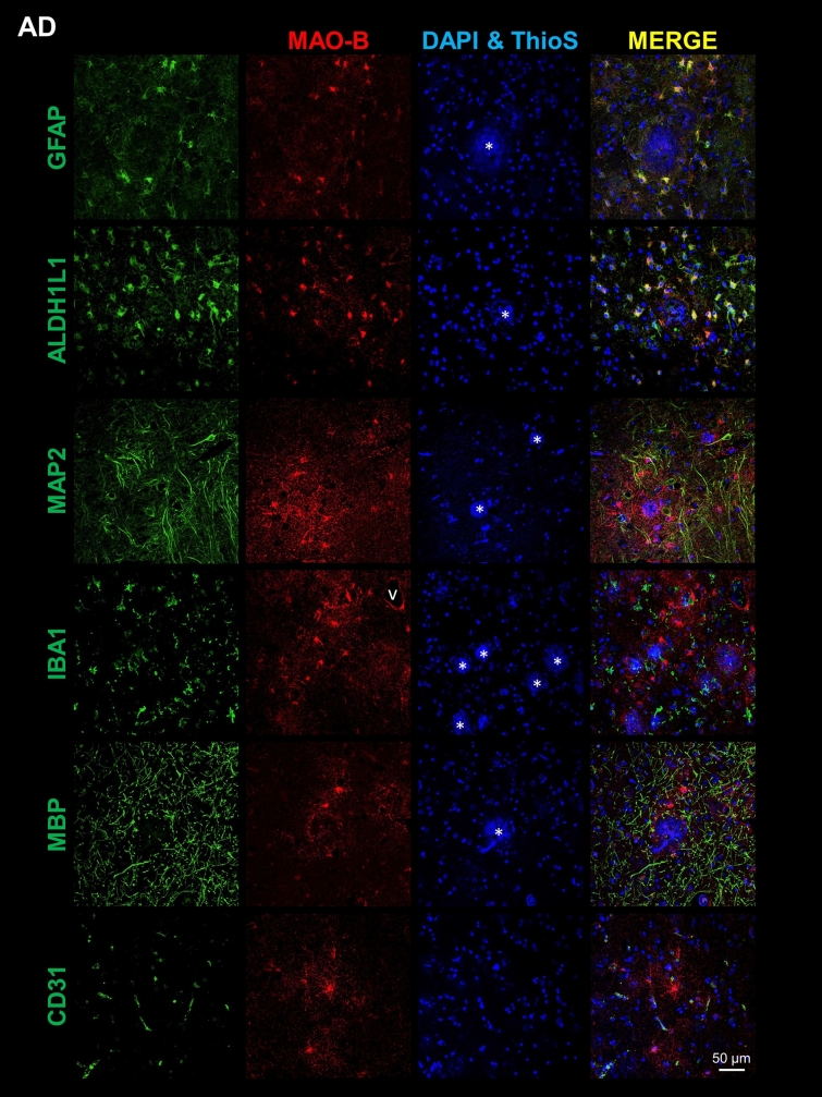
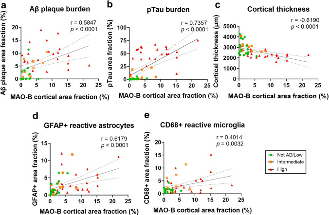
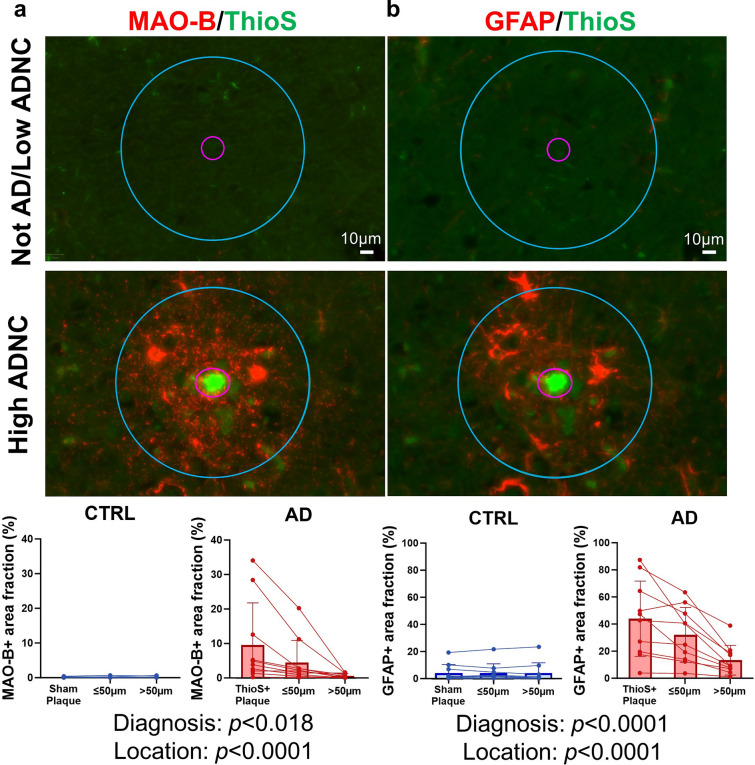
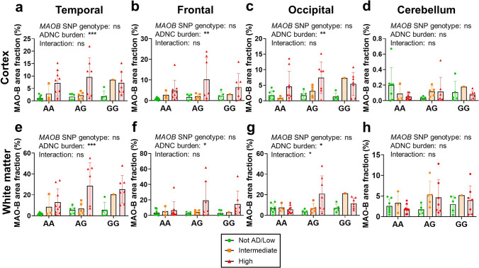
Reactive astrogliosis accompanies the two neuropathological hallmarks of Alzheimer's disease (AD)-Aβ plaques and neurofibrillary tangles-and parallels neurodegeneration in AD and AD-related dementias (ADRD). Thus, there is growing interest in developing imaging and fluid biomarkers of reactive astrogliosis for AD/ADRD diagnosis and prognostication. Monoamine oxidase-B (MAO-B) is emerging as a target for PET imaging radiotracers of reactive astrogliosis. However, a thorough characterization of MAO-B expression in postmortem control and AD/ADRD brains is lacking. We sought to: (1) identify the primary cell type(s) expressing MAO-B in control and AD brains; (2) quantify MAO-B immunoreactivity in multiple brain regions of control and AD donors as a proxy for PET radiotracer uptake; (3) correlate MAO-B level with local AD neuropathological changes, reactive glia, and cortical atrophy; (4) determine whether the MAOB rs1799836 SNP genotype impacts MAO-B expression level; (5) compare MAO-B immunoreactivity across AD/ADRD, including Lewy body diseases (LBD) and frontotemporal lobar degenerations with tau (FTLD-Tau) and TDP-43 (FTLD-TDP). We found that MAO-B is mainly expressed by subpial and perivascular cortical astrocytes as well as by fibrous white matter astrocytes in control brains, whereas in AD brains, MAO-B is significantly upregulated by both cortical reactive astrocytes and white matter astrocytes across temporal, frontal, and occipital lobes. By contrast, MAO-B expression level was unchanged and lowest in cerebellum. Cortical MAO-B expression was independently associated with cortical atrophy and local measures of reactive astrocytes and microglia, and significantly increased in reactive astrocytes surrounding Thioflavin-S+ dense-core Aβ plaques. MAO-B expression was not affected by the MAOB rs1799836 SNP genotype. MAO-B expression was also significantly increased in the frontal cortex and white matter of donors with corticobasal degeneration, Pick's disease, and FTLD-TDP, but not in LBD or progressive supranuclear palsy. These findings support ongoing efforts to develop MAO-B-based PET radiotracers to image reactive astrogliosis in AD/ADRD.
反应性星形胶质细胞伴随着阿尔茨海默病(AD)的两个神经病理学标志——β淀粉样斑块和神经原纤维缠结——与 AD 和 AD 相关痴呆(ADRD)中的神经退行性变平行。因此,人们越来越感兴趣地开发 AD/ADRD 诊断和预后的反应性星形胶质细胞的成像和液体生物标志物。单胺氧化酶-B(MAO-B)作为反应性星形胶质细胞 PET 成像示踪剂的靶标正在出现。然而,缺乏对死后对照和 AD/ADRD 大脑中 MAO-B 表达的全面表征。我们试图:(1)确定控制和 AD 大脑中表达 MAO-B 的主要细胞类型;(2)量化对照和 AD 供体多个脑区的 MAO-B 免疫反应性作为 PET 示踪剂摄取的替代物;(3)将 MAO-B 水平与局部 AD 神经病理学变化、反应性胶质和皮质萎缩相关联;(4)确定 MAOB rs1799836 SNP 基因型是否影响 MAO-B 表达水平;(5)比较 AD/ADRD 之间的 MAO-B 免疫反应性,包括路易体疾病(LBD)和额颞叶变性伴 tau(FTLD-Tau)和 TDP-43(FTLD-TDP)。我们发现 MAO-B 主要由皮质下和血管周围皮质星形胶质细胞以及纤维性白质星形胶质细胞表达,而在 AD 大脑中,MAO-B 由皮质反应性星形胶质细胞和白质星形胶质细胞显著上调,跨越颞叶、额叶和枕叶。相比之下,MAO-B 表达水平在小脑不变且最低。皮质 MAO-B 表达与皮质萎缩以及局部反应性星形胶质细胞和小胶质细胞的测量值独立相关,并且在 Thioflavin-S+致密核心 Aβ斑块周围的反应性星形胶质细胞中显著增加。MAO-B 表达不受 MAOB rs1799836 SNP 基因型的影响。MAO-B 表达在前扣带回皮质和白质中也显着增加,在皮质基底变性、皮克病和 FTLD-TDP 的供体中,但不在路易体疾病或进行性核上性麻痹中。这些发现支持正在进行的努力,以开发基于 MAO-B 的 PET 示踪剂来成像 AD/ADRD 中的反应性星形胶质细胞。