Harada Ryuichi, Furumoto Shozo, Kudo Yukitsuka, Yanai Kazuhiko, Villemagne Victor L, Okamura Nobuyuki
Department of Pharmacology, Tohoku University Graduate School of Medicine, Sendai, Japan.
Cyclotron and Radioisotope Center, Tohoku University, Sendai, Japan.
Front Neurosci. 2022 Feb 8;16:807435. doi: 10.3389/fnins.2022.807435. eCollection 2022.
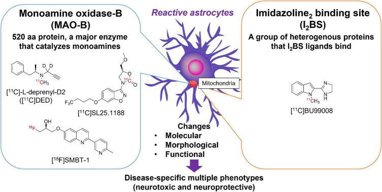
Many neurodegenerative diseases are neuropathologically characterized by neuronal loss, gliosis, and the deposition of misfolded proteins such as β-amyloid (Aβ) plaques and tau tangles in Alzheimer's disease (AD). In postmortem AD brains, reactive astrocytes and activated microglia are observed surrounding Aβ plaques and tau tangles. These activated glial cells secrete pro-inflammatory cytokines and reactive oxygen species, which may contribute to neurodegeneration. Therefore, imaging of glial response by positron emission tomography (PET) combined with Aβ and tau PET would provide new insights to better understand the disease process, as well as aid in the differential diagnosis, and monitoring glial response disease-specific therapeutics. There are two promising targets proposed for imaging reactive astrogliosis: monoamine oxidase-B (MAO-B) and imidazoline binding site (IBS), which are predominantly expressed in the mitochondrial membranes of astrocytes and are upregulated in various neurodegenerative conditions. PET tracers targeting these two MAO-B and IBS have been evaluated in humans. [F]THK-5351, which was originally designed to target tau aggregates in AD, showed high affinity for MAO-B and clearly visualized reactive astrocytes in progressive supranuclear palsy (PSP). However, the lack of selectivity of [F]THK-5351 binding to both MAO-B and tau, severely limits its clinical utility as a biomarker. Recently, [F]SMBT-1 was developed as a selective and reversible MAO-B PET tracer via compound optimization of [F]THK-5351. In this review, we summarize the strategy underlying molecular imaging of reactive astrogliosis and clinical studies using MAO-B and IBS PET tracers.
许多神经退行性疾病在神经病理学上的特征是神经元丢失、胶质细胞增生,以及在阿尔茨海默病(AD)中错误折叠蛋白的沉积,如β-淀粉样蛋白(Aβ)斑块和tau缠结。在AD患者的尸检大脑中,可观察到反应性星形胶质细胞和活化的小胶质细胞围绕着Aβ斑块和tau缠结。这些活化的胶质细胞分泌促炎细胞因子和活性氧,这可能导致神经退行性变。因此,通过正电子发射断层扫描(PET)结合Aβ和tau PET对胶质细胞反应进行成像,将为更好地理解疾病过程提供新的见解,并有助于鉴别诊断以及监测针对疾病特异性治疗的胶质细胞反应。目前提出了两个用于成像反应性星形胶质细胞增生的有前景的靶点:单胺氧化酶-B(MAO-B)和咪唑啉结合位点(IBS),它们主要表达于星形胶质细胞的线粒体膜上,并在各种神经退行性疾病状态下上调。针对这两个MAO-B和IBS的PET示踪剂已在人体中进行了评估。最初设计用于靶向AD中tau聚集体的[F]THK-5351,对MAO-B具有高亲和力,并在进行性核上性麻痹(PSP)中清晰地显示了反应性星形胶质细胞。然而,[F]THK-5351与MAO-B和tau结合均缺乏选择性,严重限制了其作为生物标志物的临床应用。最近,通过对[F]THK-5351进行化合物优化,开发出了[F]SMBT-1作为一种选择性和可逆的MAO-B PET示踪剂。在这篇综述中,我们总结了反应性星形胶质细胞增生分子成像的潜在策略以及使用MAO-B和IBS PET示踪剂的临床研究。