Medical Imaging Center, The First Affiliated Hospital of Jinan University, No. 613 West Huangpu Avenue, Tianhe District, Guangzhou, 510630, China.
UItrasonic Department, The First Affiliated Hospital of Jinan University, Guangzhou, China.
BMC Musculoskelet Disord. 2024 Apr 15;25(1):292. doi: 10.1186/s12891-024-07375-4.
Magnetic resonance imaging (MRI) can diagnose meniscal lesions anatomically, while quantitative MRI can reflect the changes of meniscal histology and biochemical structure. Our study aims to explore the association between the measurement values obtained from synthetic magnetic resonance imaging (SyMRI) and Stoller grades. Additionally, we aim to assess the diagnostic accuracy of SyMRI in determining the extent of meniscus injury. This potential accuracy could contribute to minimizing unnecessary invasive examinations and providing guidance for clinical treatment.
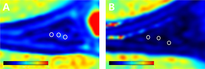
Total of 60 (n=60) patients requiring knee arthroscopic surgery and 20 (n=20) healthy subjects were collected from July 2022 to November 2022. All subjects underwent conventional MRI and SyMRI. Manual measurements of the T1, T2 and proton density (PD) values were conducted for both normal menisci and the most severely affected position of injured menisci. These measurements corresponded to the Stoller grade of meniscus injuries observed in the conventional MRI. All patients and healthy subjects were divided into normal group, degeneration group and torn group according to the Stoller grade on conventional MRI. One-way analysis of variance (ANOVA) was employed to compare the T1, T2 and PD values of the meniscus among 3 groups. The accuracy of SyMRI in diagnosing meniscus injury was assessed by comparing the findings with arthroscopic observations. The diagnostic efficiency of meniscus degeneration and tear between conventional MRI and SyMRI were analyzed using McNemar test. Furthermore, a receiver operating characteristic curve (ROC curve) was constructed and the area under the curve (AUC) was utilized for evaluation.
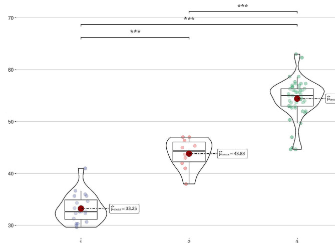
According to the measurements of SyMRI, there was no statistical difference of T1 value or PD value measured by SyMRI among the normal group, degeneration group and torn group, while the difference of T2 value was statistically significant among 3 groups (P=0.001). The arthroscopic findings showed that 11 patients were meniscal degeneration and 49 patients were meniscal tears. The arthroscopic findings were used as the gold standard, and the difference of T1 and PD values among the 3 groups was not statistically significant, while the difference of T2 values (32.81±2.51 of normal group, 44.85±3.98 of degeneration group and 54.42±3.82 of torn group) was statistically significant (P=0.001). When the threshold of T2 value was 51.67 (ms), the maximum Yoden index was 0.787 and the AUC value was 0.934.
The measurement values derived from SyMRI could reflect the Stoller grade, illustrating that SyMRI has good consistency with conventional MRI. Moreover, the notable consistency observed between SyMRI and arthroscopy suggests a potential role for SyMRI in guiding clinical diagnoses.
磁共振成像(MRI)可以从解剖学上诊断半月板损伤,而定量 MRI 可以反映半月板组织学和生化结构的变化。本研究旨在探讨合成磁共振成像(SyMRI)测量值与斯托勒(Stoller)分级之间的关系,同时评估 SyMRI 诊断半月板损伤程度的准确性。这种潜在的准确性有助于减少不必要的有创检查,并为临床治疗提供指导。
2022 年 7 月至 2022 年 11 月,共收集了 60 例(n=60)需要膝关节关节镜手术的患者和 20 例(n=20)健康受试者。所有受试者均行常规 MRI 和 SyMRI 检查。对正常半月板和损伤半月板中受影响最严重的部位进行 T1、T2 和质子密度(PD)值的手动测量,这些测量与常规 MRI 观察到的半月板斯托勒分级相对应。所有患者和健康受试者均根据常规 MRI 上的斯托勒分级分为正常组、退变组和撕裂组。采用单因素方差分析(ANOVA)比较 3 组半月板的 T1、T2 和 PD 值。通过与关节镜观察结果进行比较,评估 SyMRI 诊断半月板损伤的准确性。采用 McNemar 检验分析常规 MRI 和 SyMRI 诊断半月板退变和撕裂的诊断效率。此外,构建受试者工作特征曲线(ROC 曲线)并利用曲线下面积(AUC)进行评估。
根据 SyMRI 测量结果,正常组、退变组和撕裂组之间的 T1 值或 PD 值测量值无统计学差异,而 T2 值的差异有统计学意义(P=0.001)。关节镜检查发现 11 例半月板退变,49 例半月板撕裂。以关节镜检查结果为金标准,3 组 T1 和 PD 值差异无统计学意义,而 T2 值差异有统计学意义(正常组 32.81±2.51、退变组 44.85±3.98、撕裂组 54.42±3.82)(P=0.001)。当 T2 值的阈值为 51.67(ms)时,最大 Yoden 指数为 0.787,AUC 值为 0.934。
SyMRI 测量值可以反映斯托勒分级,表明 SyMRI 与常规 MRI 具有良好的一致性。此外,SyMRI 与关节镜检查之间的显著一致性表明,SyMRI 可能在指导临床诊断方面具有潜在作用。