Cilia Brandon-Joe, Eratne Dhamidhu, Wannan Cassandra, Malpas Charles, Janelidze Shorena, Hansson Oskar, Everall Ian, Bousman Chad, Thomas Naveen, Santillo Alexander F, Velakoulis Dennis, Pantelis Christos
Neuropsychiatry, The Royal Melbourne Hospital, Parkville, VIC, Australia.
Melbourne Medical School, The University of Melbourne, Parkville, VIC, Australia.
medRxiv. 2024 Apr 8:2024.04.07.24305362. doi: 10.1101/2024.04.07.24305362.
Around 30% of people with schizophrenia are refractory to antipsychotic treatment (treatment-resistant schizophrenia; TRS). While abnormal structural neuroimaging findings, in particular volume and thickness reductions, are often observed in schizophrenia, it is anticipated that biomarkers of neuronal injury like neurofilament light chain protein (NfL) can improve our understanding of the pathological basis underlying schizophrenia. The current study aimed to determine whether people with TRS demonstrate different associations between plasma NfL levels and regional cortical thickness reductions compared with controls.
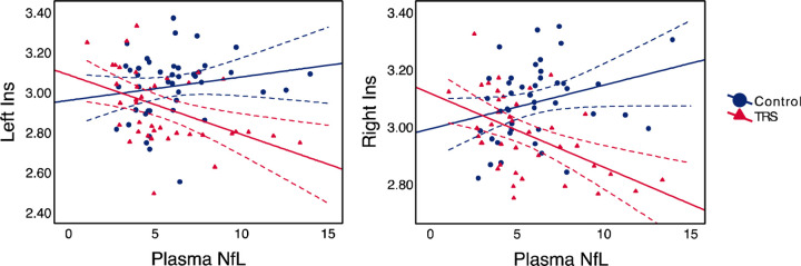
Measurements of plasma NfL and cortical thickness were obtained from 39 individuals with TRS, and 43 healthy controls. T1-weighted magnetic resonance imaging sequences were obtained and processed via FreeSurfer. General linear mixed models adjusting for age and weight were estimated to determine whether the interaction between diagnostic group and plasma NfL level predicted lower cortical thickness across frontotemporal structures and the insula.
Significant (false discovery rate corrected) cortical thinning of the left ( = 0.001, = 0.104) and right ( < 0.001, = 0.167) insula was associated with higher levels of plasma NfL in TRS, but not in healthy controls.
The association between regional thickness reduction of the insula bilaterally and plasma NfL may reflect a neurodegenerative process during the course of TRS. The findings of the present study suggest that some level of cortical degeneration localised to the bilateral insula may exist in people with TRS, which is not observed in the normal population.
约30%的精神分裂症患者对抗精神病药物治疗无效(难治性精神分裂症;TRS)。虽然在精神分裂症患者中经常观察到异常的结构神经影像学表现,特别是脑容量和厚度的减少,但预计神经元损伤生物标志物如神经丝轻链蛋白(NfL)能增进我们对精神分裂症病理基础的理解。本研究旨在确定与对照组相比,TRS患者血浆NfL水平与区域皮质厚度减少之间是否存在不同的关联。
对39例TRS患者和43名健康对照者进行血浆NfL和皮质厚度测量。通过FreeSurfer获取并处理T1加权磁共振成像序列。估计调整年龄和体重的一般线性混合模型,以确定诊断组与血浆NfL水平之间的相互作用是否能预测额颞叶结构和脑岛的皮质厚度降低。
TRS患者中,左侧脑岛( = 0.001, = 0.104)和右侧脑岛( < 0.001, = 0.167)显著(经错误发现率校正)变薄与血浆NfL水平升高相关,但在健康对照者中无此关联。
双侧脑岛区域厚度减少与血浆NfL之间的关联可能反映了TRS病程中的神经退行性过程。本研究结果表明,TRS患者可能存在某种程度的双侧脑岛局部皮质退化,而在正常人群中未观察到这种情况。