• 文献检索
  • 文档翻译
  • 深度研究
  • 学术资讯
  • Suppr Zotero 插件Zotero 插件
  • 邀请有礼
  • 套餐&价格
  • 历史记录
应用&插件
Suppr Zotero 插件Zotero 插件浏览器插件Mac 客户端Windows 客户端微信小程序
定价
高级版会员购买积分包购买API积分包
服务
文献检索文档翻译深度研究API 文档MCP 服务
关于我们
关于 Suppr公司介绍联系我们用户协议隐私条款
关注我们

Suppr 超能文献

核心技术专利:CN118964589B侵权必究
粤ICP备2023148730 号-1Suppr @ 2026

文献检索

告别复杂PubMed语法,用中文像聊天一样搜索,搜遍4000万医学文献。AI智能推荐,让科研检索更轻松。

立即免费搜索

文件翻译

保留排版,准确专业,支持PDF/Word/PPT等文件格式,支持 12+语言互译。

免费翻译文档

深度研究

AI帮你快速写综述,25分钟生成高质量综述,智能提取关键信息,辅助科研写作。

立即免费体验

通过控制射线照相体模的欠填充来调整3D打印材料的质量密度以进行精确的X射线成像模拟

Tailoring the Mass Density of 3D Printing Materials for Accurate X-ray Imaging Simulation by Controlled Underfilling for Radiographic Phantoms.

作者信息

Ahmed Ahmed Mahmoud Mabrouk, Buschmann Martin, Breyer Lara, Kuntner Claudia, Homolka Peter

机构信息

Center for Medical Physics and Biomedical Engineering, Medical University of Vienna, 1090 Vienna, Austria.

Division of Medical Radiation Physics, Department of Radiation Oncology, Medical University of Vienna, and University Hospital Vienna, 1090 Vienna, Austria.

出版信息

Polymers (Basel). 2024 Apr 16;16(8):1116. doi: 10.3390/polym16081116.

DOI:10.3390/polym16081116
PMID:38675035
原文链接:https://pmc.ncbi.nlm.nih.gov/articles/PMC11053449/
Abstract

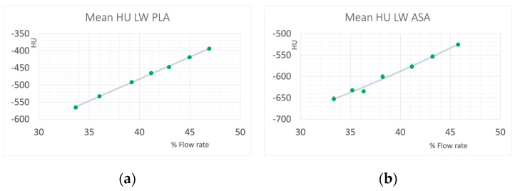
Additive manufacturing and 3D printing allow for the design and rapid production of radiographic phantoms for X-ray imaging, including CT. These are used for numerous purposes, such as patient simulation, optimization of imaging procedures and dose levels, system evaluation and quality assurance. However, standard 3D printing polymers do not mimic X-ray attenuation properties of tissues like soft, adipose, lung or bone tissue, and standard materials like liquid water. The mass density of printing polymers-especially important in CT-is often inappropriate, i.e., mostly too high. Different methods can be applied to reduce mass density. This work examines reducing density by controlled underfilling either realized by using 3D printing materials expanded through foaming during heating in the printing process, or reducing polymer flow to introduce microscopic air-filled voids. The achievable density reduction depends on the base polymer used. When using foaming materials, density is controlled by the extrusion temperature, and ranges from 33 to 47% of the base polymer used, corresponding to a range of -650 to -394 HU in CT with 120 kV. Standard filaments (Nylon, modified PLA and modified ABS) allowed density reductions by 20 to 25%, covering HU values in CT from -260 to 77 (Nylon), -230 to -20 (ABS) and -81 to 143 (PLA). A standard chalk-filled PLA filament allowed reproduction of bone tissue in a wide range of bone mineral content resulting in CT numbers from 57 to 460 HU. Controlled underfilling allowed the production of radiographic phantom materials with continuously adjustable attenuation in a limited but appropriate range, allowing for the reproduction of X-ray attenuation properties of water, adipose, soft, lung, and bone tissue in an accurate, predictable and reproducible manner.

摘要

增材制造和3D打印技术可用于设计和快速生产用于X射线成像(包括CT)的放射造影体模。这些体模有多种用途,如患者模拟、成像程序和剂量水平的优化、系统评估和质量保证。然而,标准的3D打印聚合物无法模拟软组织、脂肪组织、肺组织或骨组织等组织以及液态水等标准材料的X射线衰减特性。打印聚合物的质量密度(在CT中尤为重要)通常不合适,即大多过高。可以采用不同方法来降低质量密度。本研究探讨了通过控制欠填充来降低密度的方法,具体实现方式要么是使用在打印过程中加热时通过发泡膨胀的3D打印材料,要么是减少聚合物流动以引入微观的空气填充空隙。可实现的密度降低程度取决于所使用的基础聚合物。使用发泡材料时,密度由挤出温度控制,范围为所用基础聚合物的33%至47%,在120 kV的CT中对应-650至-394 HU的范围。标准细丝(尼龙、改性聚乳酸和改性丙烯腈-丁二烯-苯乙烯)可使密度降低20%至25%,在CT中的HU值范围为-260至77(尼龙)、-230至-20(丙烯腈-丁二烯-苯乙烯)和-81至143(聚乳酸)。一种标准的填充白垩聚乳酸细丝能够在广泛的骨矿物质含量范围内再现骨组织,CT值范围为57至460 HU。控制欠填充能够生产出在有限但合适范围内具有连续可调衰减的放射造影体模材料,从而能够以准确、可预测和可重复的方式再现水、脂肪、软组织、肺组织和骨组织的X射线衰减特性。

https://cdn.ncbi.nlm.nih.gov/pmc/blobs/1a29/11053449/4418e9add238/polymers-16-01116-g007.jpg
https://cdn.ncbi.nlm.nih.gov/pmc/blobs/1a29/11053449/9d03697edb12/polymers-16-01116-g001.jpg
https://cdn.ncbi.nlm.nih.gov/pmc/blobs/1a29/11053449/05a3f7d418be/polymers-16-01116-g002.jpg
https://cdn.ncbi.nlm.nih.gov/pmc/blobs/1a29/11053449/bd4f497e46d2/polymers-16-01116-g003.jpg
https://cdn.ncbi.nlm.nih.gov/pmc/blobs/1a29/11053449/be930496f13f/polymers-16-01116-g004.jpg
https://cdn.ncbi.nlm.nih.gov/pmc/blobs/1a29/11053449/79f9d5d9cabf/polymers-16-01116-g005.jpg
https://cdn.ncbi.nlm.nih.gov/pmc/blobs/1a29/11053449/3a637f606c4b/polymers-16-01116-g006.jpg
https://cdn.ncbi.nlm.nih.gov/pmc/blobs/1a29/11053449/4418e9add238/polymers-16-01116-g007.jpg

相似文献

1
Tailoring the Mass Density of 3D Printing Materials for Accurate X-ray Imaging Simulation by Controlled Underfilling for Radiographic Phantoms.通过控制射线照相体模的欠填充来调整3D打印材料的质量密度以进行精确的X射线成像模拟
Polymers (Basel). 2024 Apr 16;16(8):1116. doi: 10.3390/polym16081116.
2
Classification of X-Ray Attenuation Properties of Additive Manufacturing and 3D Printing Materials Using Computed Tomography From 70 to 140 kVp.使用70至140 kVp的计算机断层扫描对增材制造和3D打印材料的X射线衰减特性进行分类
Front Bioeng Biotechnol. 2021 Nov 29;9:763960. doi: 10.3389/fbioe.2021.763960. eCollection 2021.
3
X-ray attenuation of bone, soft and adipose tissue in CT from 70 to 140 kV and comparison with 3D printable additive manufacturing materials.CT 从 70 到 140kV 时骨、软组织和脂肪组织的 X 射线衰减及与 3D 打印增材制造材料的比较。
Sci Rep. 2022 Aug 26;12(1):14580. doi: 10.1038/s41598-022-18741-4.
4
A voxel-by-voxel method for mixing two filaments during a 3D printing process for soft-tissue replication in an anthropomorphic breast phantom.一种用于在人体乳房模型中进行软组织复制的 3D 打印过程中混合两条纤维的体素对体素方法。
Phys Med Biol. 2022 Dec 15;67(24). doi: 10.1088/1361-6560/aca640.
5
3D printed patient-specific thorax phantom with realistic heterogenous bone radiopacity using filament printer technology.使用丝材挤出式 3D 打印机技术打印具有真实异质骨不透明度的个体化胸腔体模。
Z Med Phys. 2022 Nov;32(4):438-452. doi: 10.1016/j.zemedi.2022.02.001. Epub 2022 Feb 24.
6
3D Printing Materials Mimicking Human Tissues after Uptake of Iodinated Contrast Agents for Anthropomorphic Radiology Phantoms.用于拟人化放射学体模的摄取碘化造影剂后模仿人体组织的3D打印材料。
Biomimetics (Basel). 2024 Oct 8;9(10):606. doi: 10.3390/biomimetics9100606.
7
A filament 3D printing approach for CT-compatible bone tissues replication.一种用于复制 CT 兼容骨组织的细丝 3D 打印方法。
Phys Med. 2022 Oct;102:96-102. doi: 10.1016/j.ejmp.2022.09.009. Epub 2022 Sep 23.
8
A customizable anthropomorphic phantom for dosimetric verification of 3D-printed lung, tissue, and bone density materials.一种可定制的拟人化体模,用于对 3D 打印的肺、组织和骨密度材料进行剂量学验证。
Med Phys. 2022 Jan;49(1):52-69. doi: 10.1002/mp.15364. Epub 2021 Dec 2.
9
Fabrication of a pediatric torso phantom with multiple tissues represented using a dual nozzle thermoplastic 3D printer.使用双喷嘴热塑性3D打印机制造具有多种组织的儿科躯干模型。
J Appl Clin Med Phys. 2020 Nov;21(11):226-236. doi: 10.1002/acm2.13064. Epub 2020 Oct 19.
10
Methodology for computed tomography characterization of commercially available 3D printing materials for use in radiology/radiation oncology.用于放射学/放射肿瘤学的商用 3D 打印材料的计算机断层扫描特征化方法。
J Appl Clin Med Phys. 2023 Jun;24(6):e13999. doi: 10.1002/acm2.13999. Epub 2023 Apr 24.

引用本文的文献

1
Fast and Fractionated: Correlation of Dose Attenuation and the Response of Human Cancer Cells in a New Anthropomorphic Brain Phantom.快速与分次:在新型人体头部仿真体模中剂量衰减与人类癌细胞反应的相关性
Biomimetics (Basel). 2025 Jul 3;10(7):440. doi: 10.3390/biomimetics10070440.
2
3D Printing Materials Mimicking Human Tissues after Uptake of Iodinated Contrast Agents for Anthropomorphic Radiology Phantoms.用于拟人化放射学体模的摄取碘化造影剂后模仿人体组织的3D打印材料。
Biomimetics (Basel). 2024 Oct 8;9(10):606. doi: 10.3390/biomimetics9100606.

本文引用的文献

1
Characterization of 3-Dimensional Printing and Casting Materials for use in Computed Tomography and X-ray Imaging Phantoms.用于计算机断层扫描和X射线成像体模的三维打印及铸造材料的特性研究
J Res Natl Inst Stand Technol. 2020 Sep 15;125:125029. doi: 10.6028/jres.125.029. eCollection 2020.
2
X-ray attenuation of bone, soft and adipose tissue in CT from 70 to 140 kV and comparison with 3D printable additive manufacturing materials.CT 从 70 到 140kV 时骨、软组织和脂肪组织的 X 射线衰减及与 3D 打印增材制造材料的比较。
Sci Rep. 2022 Aug 26;12(1):14580. doi: 10.1038/s41598-022-18741-4.
3
Characterization of 3-Dimensional Printing and Casting Materials for use in Magnetic Resonance Imaging Phantoms at 3 T.
用于3T磁共振成像体模的三维打印及铸造材料的特性研究
J Res Natl Inst Stand Technol. 2020 Sep 15;vol. doi: 10.6028/jres.125.028. eCollection 2020.
4
3D printed patient-specific thorax phantom with realistic heterogenous bone radiopacity using filament printer technology.使用丝材挤出式 3D 打印机技术打印具有真实异质骨不透明度的个体化胸腔体模。
Z Med Phys. 2022 Nov;32(4):438-452. doi: 10.1016/j.zemedi.2022.02.001. Epub 2022 Feb 24.
5
Technical note: Controlling the attenuation of 3D-printed physical phantoms for computed tomography with a single material.技术说明:使用单一材料控制 CT 中 3D 打印物理模型的衰减。
Med Phys. 2022 Apr;49(4):2582-2589. doi: 10.1002/mp.15494. Epub 2022 Mar 7.
6
Classification of X-Ray Attenuation Properties of Additive Manufacturing and 3D Printing Materials Using Computed Tomography From 70 to 140 kVp.使用70至140 kVp的计算机断层扫描对增材制造和3D打印材料的X射线衰减特性进行分类
Front Bioeng Biotechnol. 2021 Nov 29;9:763960. doi: 10.3389/fbioe.2021.763960. eCollection 2021.
7
Three-dimensional printing of patient-specific lung phantoms for CT imaging: Emulating lung tissue with accurate attenuation profiles and textures.用于 CT 成像的患者特异性肺模型的三维打印:用准确的衰减曲线和纹理模拟肺组织。
Med Phys. 2022 Feb;49(2):825-835. doi: 10.1002/mp.15407. Epub 2021 Dec 23.
8
Fabrication of a pediatric torso phantom with multiple tissues represented using a dual nozzle thermoplastic 3D printer.使用双喷嘴热塑性3D打印机制造具有多种组织的儿科躯干模型。
J Appl Clin Med Phys. 2020 Nov;21(11):226-236. doi: 10.1002/acm2.13064. Epub 2020 Oct 19.
9
3D printed soft surgical planning prototype for a biliary tract rhabdomyosarcoma.用于胆道横纹肌肉瘤的3D打印软组织手术规划原型。
J Mech Behav Biomed Mater. 2020 Sep;109:103844. doi: 10.1016/j.jmbbm.2020.103844. Epub 2020 May 11.
10
3D printing of radioactive phantoms for nuclear medicine imaging.用于核医学成像的放射性体模的3D打印。
EJNMMI Phys. 2020 Apr 22;7(1):22. doi: 10.1186/s40658-020-00292-0.