Department of Surgery, College of Medicine and Health Sciences, Gondar, Ethiopia.
Department of Pathology, College of Medicine and Health Sciences, Gondar, Ethiopia.
J Med Case Rep. 2024 Apr 29;18(1):212. doi: 10.1186/s13256-024-04503-5.
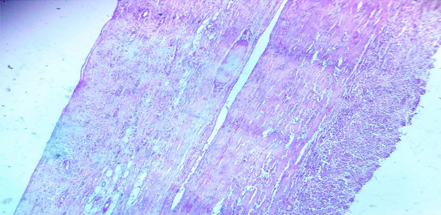
Non-pancreatic pseudocysts are rare lesions that typically form from the omentum and mesentery. These cysts have a thick fibrotic wall made up of fibrous tissue and may show signs of calcifications and inflammatory changes. The fluid inside them can vary, ranging from hemorrhage and pus to serous or sometimes chylous content. In most cases, these cysts appear as a result of trauma, surgery, or infection.
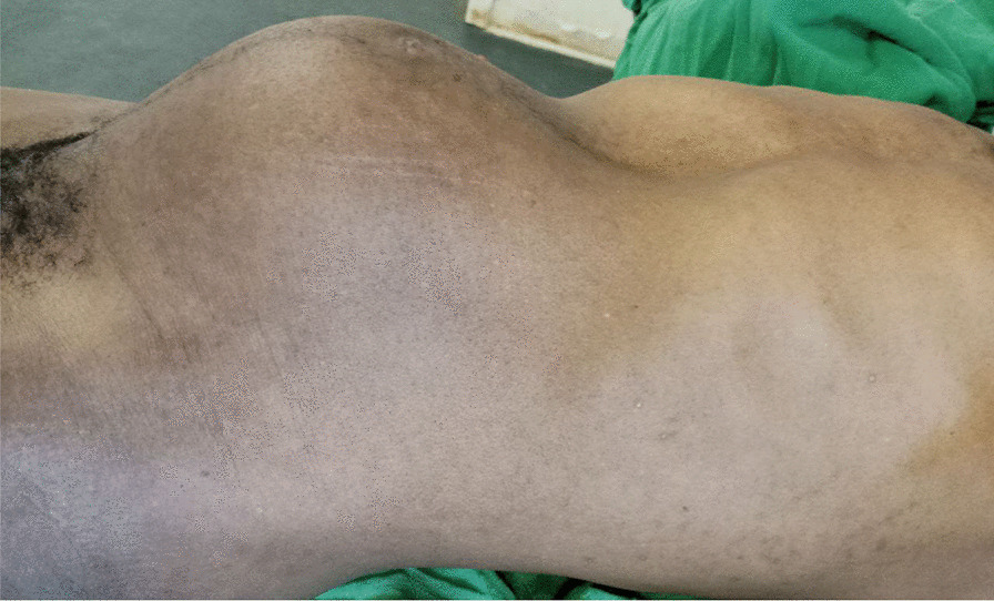
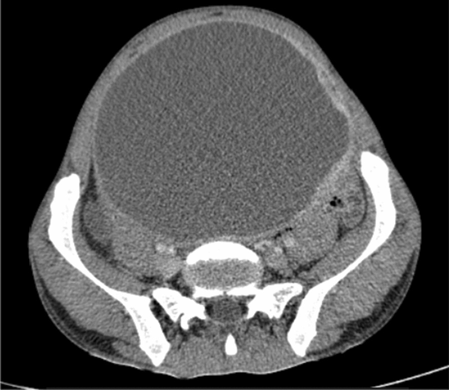
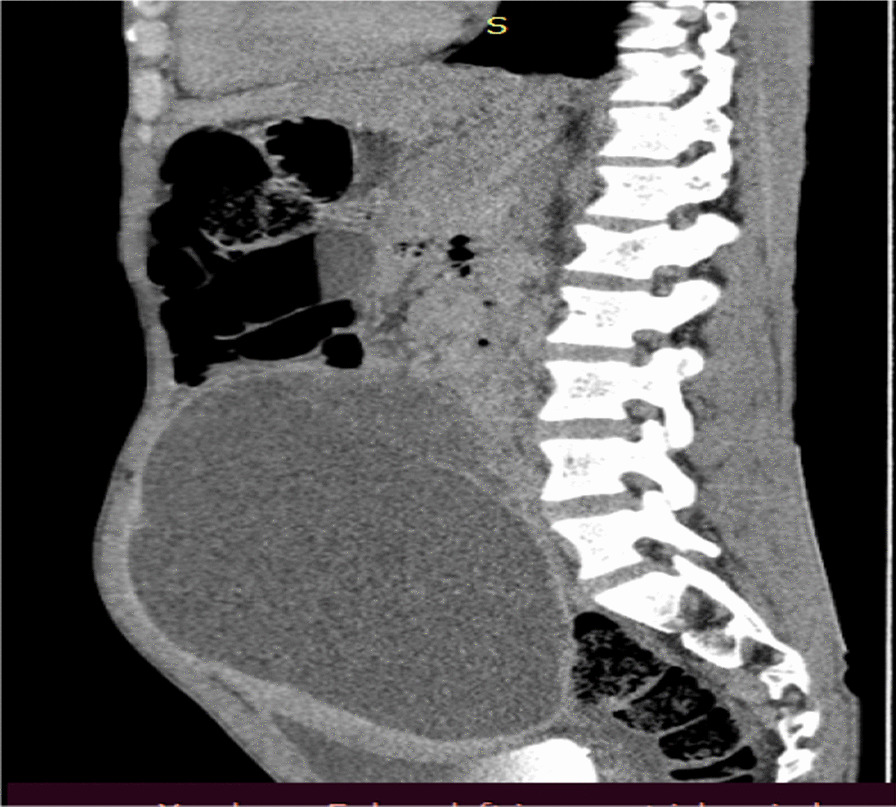
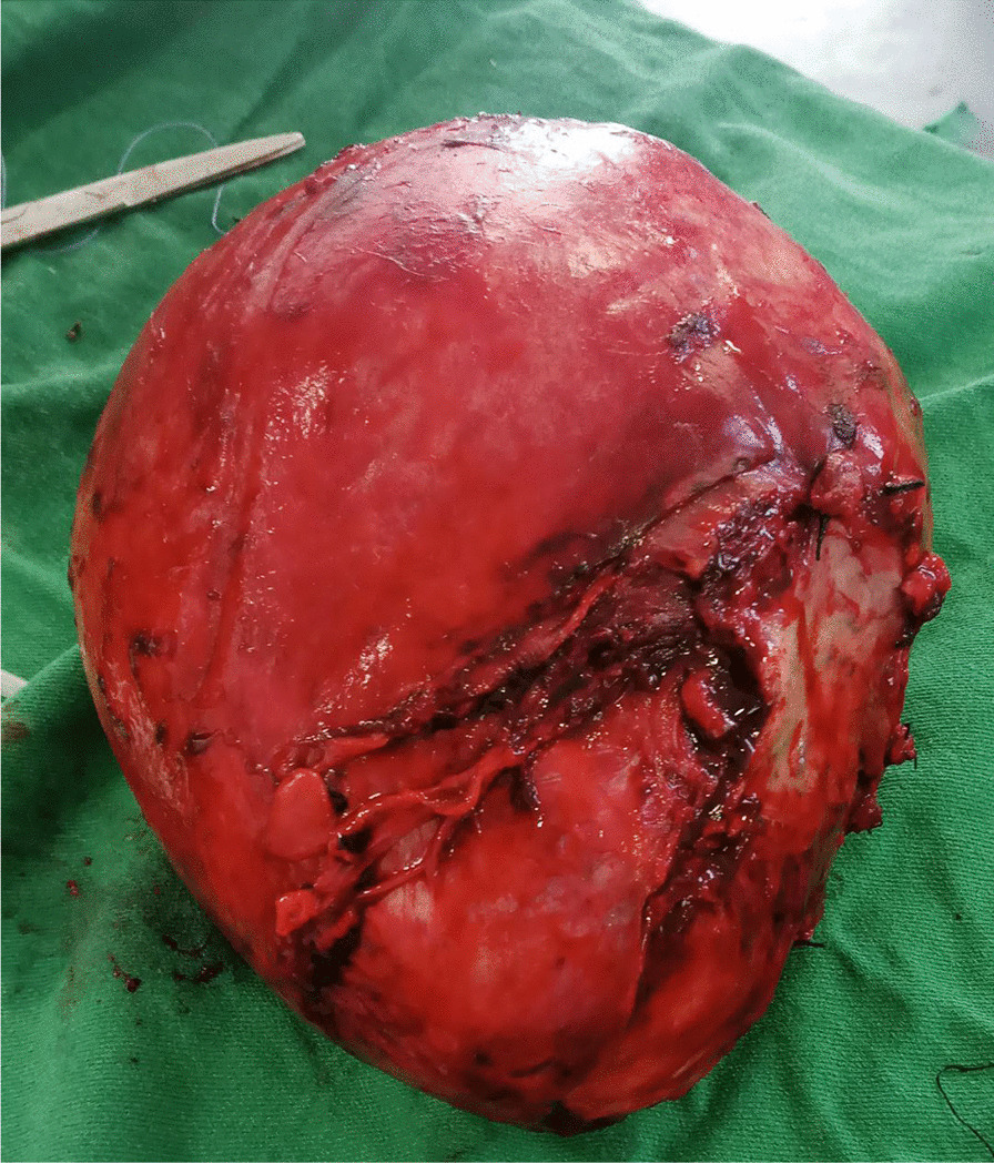
A 35-year-old male patient from Ethiopia presented with swelling in his lower abdomen that had been present for 2 years. Initially, the swelling was small but gradually increased in size. The patient experienced frequent urination but no pain or difficulty during urination, urgency, intermittent urination, or blood in the urine. The swelling was initially painless but became painful 2 months prior to his presentation. Abdominal computed tomography scans revealed a well-defined, lobulated peritoneal lesion measuring 16 × 12 × 10 cm, consisting primarily of fluid-filled cysts with a thick, enhancing wall and septa. Additionally, there was a large, heterogeneous enhancing soft tissue component measuring 8 × 6 cm. As a result, the cystic mass was surgically removed in its entirety with partial removal of the bladder wall, and the patient was discharged in an improved condition.
Primary non-pancreatic pseudocysts are extremely rare lesions that must be differentiated from other possible causes of cystic lesions within the peritoneal or retroperitoneal regions. Surgeons should be aware of the potential occurrence of these lesions, which may have an unknown origin.
非胰腺性假性囊肿是罕见的病变,通常由大网膜和肠系膜形成。这些囊肿具有由纤维组织组成的厚纤维化壁,并可能显示出钙化和炎症变化的迹象。其内部的液体可以变化,从出血和脓液到浆液性或有时乳糜性内容物。在大多数情况下,这些囊肿是由于创伤、手术或感染而出现的。
一位来自埃塞俄比亚的 35 岁男性患者,其下腹部肿胀已存在 2 年。最初,肿胀较小,但逐渐增大。患者经常排尿,但无尿痛、排尿困难、尿急、间歇性排尿或血尿。肿胀最初无痛,但在就诊前 2 个月开始疼痛。腹部计算机断层扫描显示一个界限清楚的、分叶状的腹膜病变,大小为 16×12×10cm,主要由充满液体的囊肿组成,具有厚的、增强的壁和隔膜。此外,还有一个大的、不均匀增强的软组织成分,大小为 8×6cm。因此,整个囊性肿块被手术切除,部分膀胱壁也被切除,患者出院时病情有所改善。
原发性非胰腺性假性囊肿是非常罕见的病变,必须与腹膜或腹膜后区域内其他可能导致囊性病变的原因区分开来。外科医生应该意识到这些病变的潜在发生,这些病变可能来源不明。