W. Harry Feinstone Department of Molecular Microbiology and Immunology, Johns Hopkins Bloomberg School of Public Health, Baltimore, Maryland, USA.
mBio. 2024 Jun 12;15(6):e0073624. doi: 10.1128/mbio.00736-24. Epub 2024 May 2.
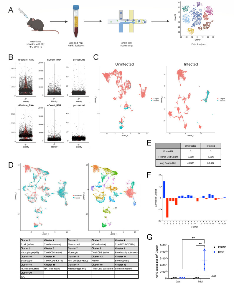
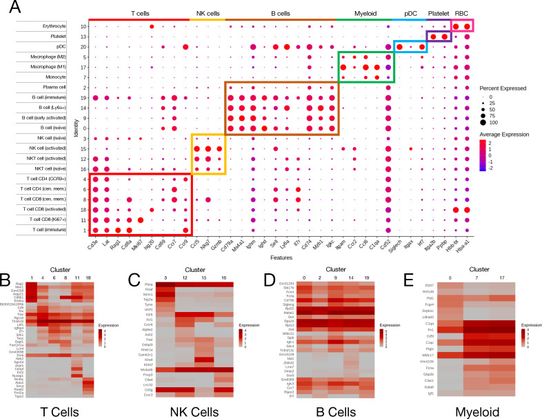
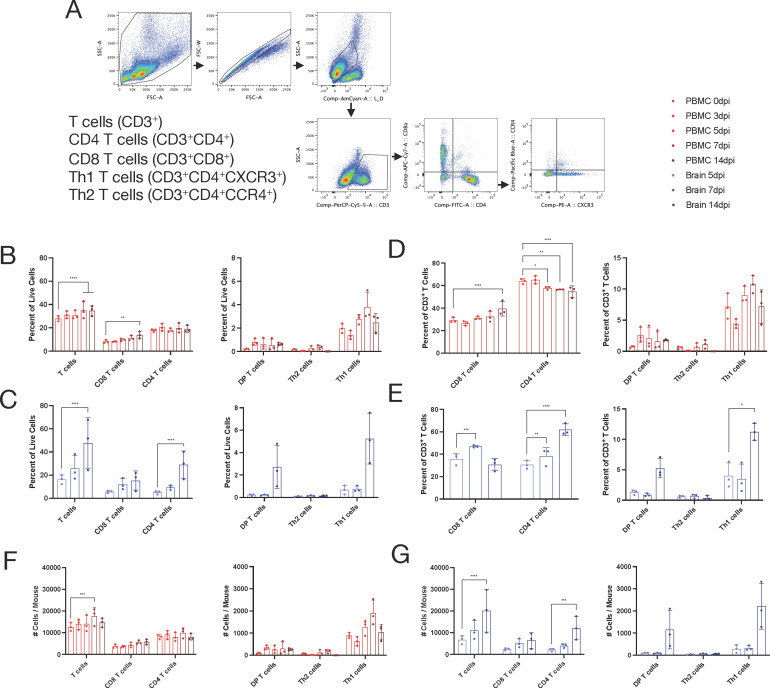
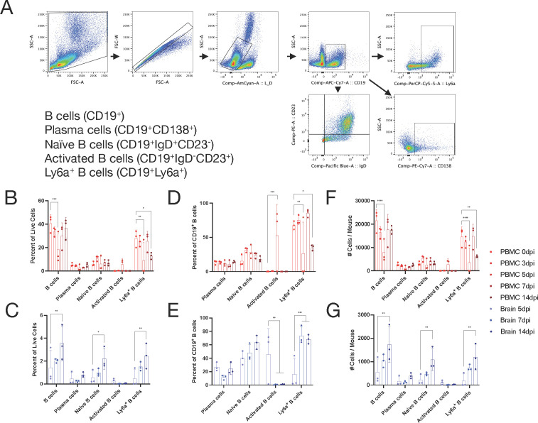
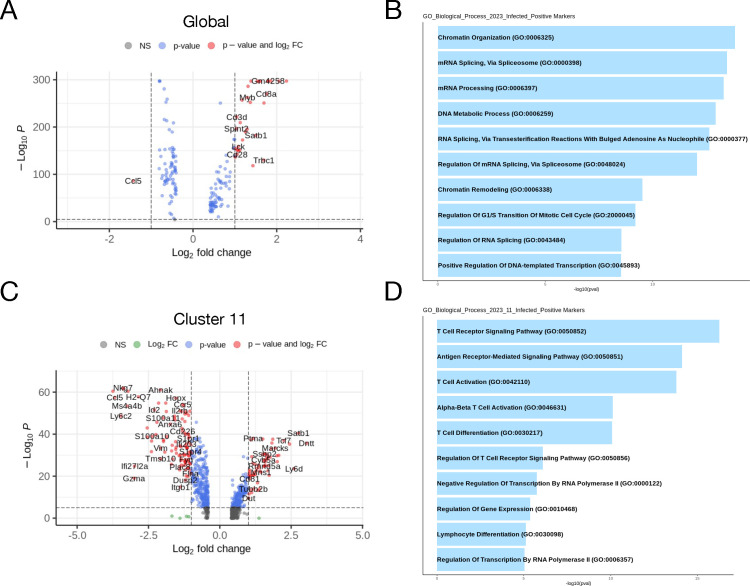
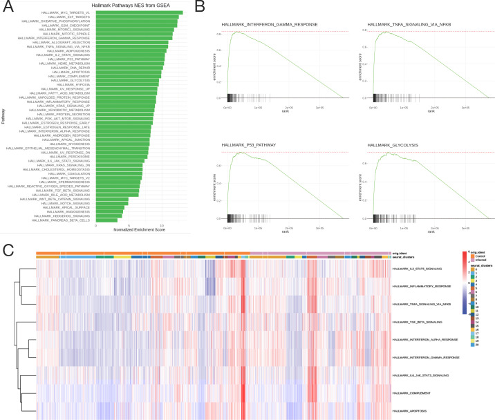
Sindbis virus (SINV) infection of mice provides a model system for studying the pathogenesis of alphaviruses that infect the central nervous system (CNS) to cause encephalomyelitis. While studies of human viral infections typically focus on accessible cells from the blood, this compartment is rarely evaluated in mice. To bridge this gap, single-cell RNA sequencing (scRNAseq) was combined with flow cytometry to characterize the transcriptional and phenotypic changes of peripheral blood mononuclear cells (PBMCs) from SINV-infected mice. Twenty-one clusters were identified by scRNAseq at 7 days after infection, with a unique cluster and overall increase in naive B cells for infected mice. Uninfected mice had fewer immature T cells and CCR9 CD4 T cells and a unique immature T cell cluster. Gene expression was most altered in the Ki67 CD8 T cell cluster, with chemotaxis and proliferation-related genes upregulated. Global analysis indicated metabolic changes in myeloid cells and increased expression of by NK cells. Phenotypes of PBMCs and cells infiltrating the CNS were analyzed by flow cytometry over 14 days after infection. In PBMCs, CD8 and Th1 CD4 T cells increased in representation, while B cells showed a transient decrease at day 5 in total, Ly6a, and naive cells, and an increase in activated B cells. In the brain, CD8 T cells increased for the first 7 days, while Th1 CD4 T cells and naive and Ly6a B cells continued to accumulate for 14 days. Therefore, dynamic immune cell changes can be identified in the blood as well as the CNS during viral encephalomyelitis.
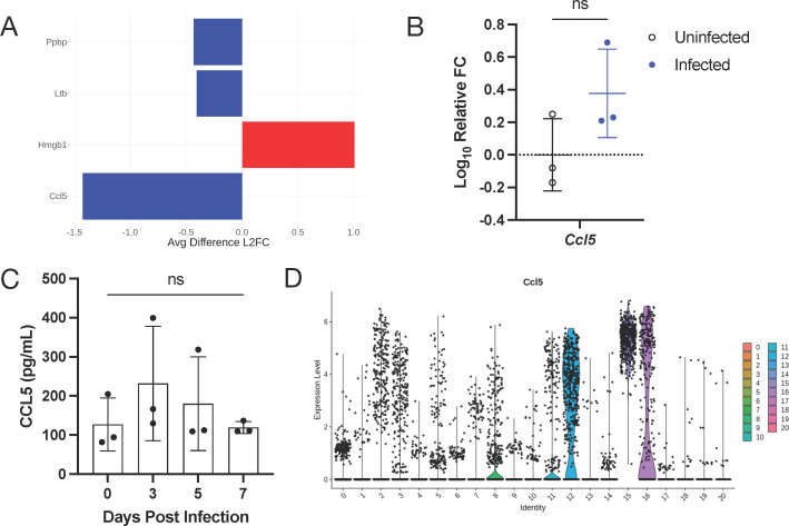
The outcome of viral encephalomyelitis is dependent on the host immune response, with clearance and resolution of infection mediated by the adaptive immune response. These processes are frequently studied in mouse models of infection, where infected tissues are examined to understand the mechanisms of clearance and recovery. However, studies of human infection typically focus on the analysis of cells from the blood, a compartment rarely examined in mice, rather than inaccessible tissue. To close this gap, we used single-cell RNA sequencing and flow cytometry to profile the transcriptomic and phenotypic changes of peripheral blood mononuclear cells (PBMCs) before and after central nervous system (CNS) infection in mice. Changes to T and B cell gene expression and cell composition occurred in PBMC and during entry into the CNS, with CCL5 being a differentially expressed chemokine. Therefore, dynamic changes occur in the blood as well as the CNS during the response of mice to virus infection, which will inform the analysis of human studies.
辛德比斯病毒(SINV)感染小鼠为研究感染中枢神经系统(CNS)引起脊髓灰质炎的甲病毒的发病机制提供了模型系统。虽然人类病毒感染的研究通常集中在血液中可获得的细胞上,但在小鼠中很少评估该隔室。为了弥补这一差距,我们将单细胞 RNA 测序(scRNAseq)与流式细胞术相结合,以描绘 SINV 感染小鼠外周血单个核细胞(PBMC)的转录和表型变化。在感染后 7 天通过 scRNAseq 鉴定出 21 个簇,感染小鼠的幼稚 B 细胞具有独特的簇和整体增加。未感染的小鼠幼稚 T 细胞和 CCR9 CD4 T 细胞较少,并且存在独特的幼稚 T 细胞簇。Ki67 CD8 T 细胞簇中的基因表达变化最大,趋化因子和增殖相关基因上调。髓样细胞的代谢变化和 NK 细胞中表达增加表明了整体分析。通过流式细胞术在感染后 14 天分析 PBMC 和浸润中枢神经系统的细胞的表型。在 PBMC 中,CD8 和 Th1 CD4 T 细胞的代表性增加,而 B 细胞的总数量,Ly6a 和幼稚细胞在第 5 天短暂减少,而激活的 B 细胞增加。在大脑中,CD8 T 细胞在最初的 7 天内增加,而 Th1 CD4 T 细胞和幼稚及 Ly6a B 细胞在 14 天内持续增加。因此,在病毒性脑脊髓炎期间,可在血液以及中枢神经系统中识别出动态的免疫细胞变化。
病毒性脑脊髓炎的结果取决于宿主免疫反应,适应性免疫反应介导了感染的清除和缓解。这些过程经常在感染的小鼠模型中进行研究,在这些模型中检查受感染的组织以了解清除和恢复的机制。但是,人类感染的研究通常集中在分析血液中的细胞上,这是在小鼠中很少检查的隔室,而不是无法进入的组织。为了弥补这一差距,我们使用单细胞 RNA 测序和流式细胞术来描绘感染前后小鼠外周血单个核细胞(PBMC)的转录组和表型变化中枢神经系统(CNS)。T 和 B 细胞基因表达和细胞组成的变化发生在 PBMC 中,并在进入中枢神经系统时发生,CCL5 是差异表达的趋化因子。因此,在小鼠对病毒感染的反应过程中,血液和中枢神经系统中均会发生动态变化,这将为人类研究的分析提供信息。