https://ror.org/05akvb491 Department of Nephrology, The Third Xiangya Hospital, Central South University, Changsha, China.
Clinical Research Center for Critical Kidney Disease in Hunan Province, Changsha, China.
Life Sci Alliance. 2024 May 2;7(7). doi: 10.26508/lsa.202302474. Print 2024 Jul.
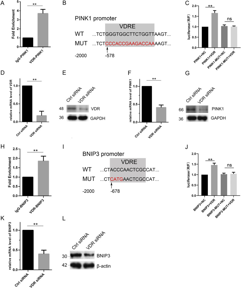
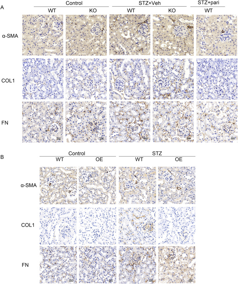
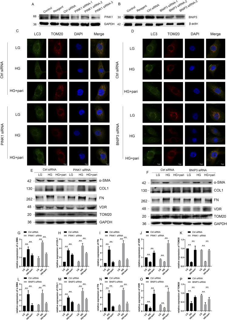
Defective mitophagy in renal tubular epithelial cells is one of the main drivers of renal fibrosis in diabetic kidney disease. Our gene sequencing data showed the expression of PINK1 and BNIP3, two key molecules of mitophagy, was decreased in renal tissues of VDR-knockout mice. Herein, streptozotocin (STZ) was used to induce renal interstitial fibrosis in mice. VDR deficiency exacerbated STZ-induced renal impairment and defective mitophagy. Paricalcitol (pari, a VDR agonist) and the tubular epithelial cell-specific overexpression of VDR restored the expression of PINK1 and BNIP3 in the renal cortex and attenuated STZ-induced kidney fibrosis and mitochondrial dysfunction. In HK-2 cells under high glucose conditions, an increased level of α-SMA, COL1, and FN and a decreased expression of PINK1 and BNIP3 with severe mitochondrial damage were observed, and these alterations could be largely reversed by pari treatment. ChIP-qPCR and luciferase reporter assays showed VDR could positively regulate the transcription of and genes. These findings reveal that VDR could restore mitophagy defects and attenuate STZ-induced fibrosis in diabetic mice through regulation of PINK1 and BNIP3.
线粒体自噬缺陷是糖尿病肾病肾小管上皮细胞肾纤维化的主要驱动因素之一。我们的基因测序数据显示,VDR 敲除小鼠的肾脏组织中,两种关键的线粒体自噬分子 PINK1 和 BNIP3 的表达减少。本文中,链脲佐菌素(STZ)用于诱导小鼠肾间质纤维化。VDR 缺乏会加重 STZ 诱导的肾损伤和线粒体自噬缺陷。帕立骨化醇(pari,一种 VDR 激动剂)和肾小管上皮细胞特异性过表达 VDR 可恢复肾脏皮质中 PINK1 和 BNIP3 的表达,并减轻 STZ 诱导的肾脏纤维化和线粒体功能障碍。在高糖条件下的 HK-2 细胞中,观察到 α-SMA、COL1 和 FN 的水平增加,PINK1 和 BNIP3 的表达减少,线粒体损伤严重,而 pari 处理可显著逆转这些改变。ChIP-qPCR 和荧光素酶报告基因检测显示 VDR 可正向调控 和 基因的转录。这些发现表明,VDR 通过调节 PINK1 和 BNIP3 可恢复糖尿病小鼠的线粒体自噬缺陷并减轻 STZ 诱导的纤维化。