Department of Biophysics, Manipal School of Life Sciences, Manipal Academy of Higher Education, Manipal, 576104, India.
Department of Instrumentation and Control Engineering, Manipal Institute of Technology, Manipal Academy of Higher Education, Manipal, Karnataka, 576104, India.
Lasers Med Sci. 2024 May 4;39(1):123. doi: 10.1007/s10103-024-04056-5.
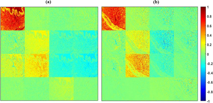
Interaction of polarized light with healthy and abnormal regions of tissue reveals structural information associated with its pathological condition. Even a slight variation in structural alignment can induce a change in polarization property, which can play a crucial role in the early detection of abnormal tissue morphology. We propose a transmission-based Stokes-Mueller microscope for quantitative analysis of the microstructural properties of the tissue specimen. The Stokes-Mueller based polarization microscopy provides significant structural information of tissue through various polarization parameters such as degree of polarization (DOP), degree of linear polarization (DOLP), and degree of circular polarization (DOCP), anisotropy (r) and Mueller decomposition parameters such as diattenuation, retardance and depolarization. Further, by applying a suitable image processing technique such as Machine learning (ML) output images were analysed effectively. The support vector machine image classification model achieved 95.78% validation accuracy and 94.81% testing accuracy with polarization parameter dataset. The study's findings demonstrate the potential of Stokes-Mueller polarimetry in tissue characterization and diagnosis, providing a valuable tool for biomedical applications.
偏振光与组织的健康和异常区域的相互作用揭示了与其病理状况相关的结构信息。即使结构排列的微小变化也会引起偏振特性的变化,这在异常组织形态的早期检测中起着至关重要的作用。我们提出了一种基于传输的斯托克斯-穆勒显微镜,用于定量分析组织标本的微观结构特性。基于斯托克斯-穆勒的偏振显微镜通过各种偏振参数(如偏振度(DOP)、线偏振度(DOLP)和圆偏振度(DOCP)、各向异性(r)和穆勒分解参数(如消光、延迟和去偏)提供组织的重要结构信息。此外,通过应用合适的图像处理技术(如机器学习(ML)),可以有效地分析输出图像。基于偏振参数数据集的支持向量机图像分类模型的验证准确率为 95.78%,测试准确率为 94.81%。该研究结果表明,斯托克斯-穆勒偏振术在组织特征化和诊断中有很大的应用潜力,为生物医学应用提供了一种有价值的工具。