Translational Virology, Department of Medical Microbiology, University Medical Center Utrecht, 3584 CX, Utrecht, The Netherlands.
Department of Internal Medicine and Infectious Diseases, University Medical Center Utrecht, 3584 CX, Utrecht, The Netherlands.
J Neurovirol. 2024 Aug;30(4):380-392. doi: 10.1007/s13365-024-01207-w. Epub 2024 May 7.
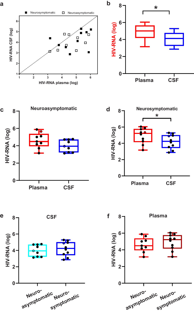
Despite antiretroviral therapy (ART), HIV persistence in the central nervous system (CNS) continues to cause a range of cognitive impairments in people living with HIV (PLWH). Upon disease progression, transmigrating CCR5-using T-cell tropic viruses are hypothesized to evolve into macrophage-tropic viruses in the CNS that can efficiently infect low CD4-expressing cells, such as microglia. We examined HIV-1 RNA concentration, co-receptor usage, and CSF compartmentalization in paired CSF and blood samples from 19 adults not on treatment. Full-length envelope CSF- and plasma-derived reporter viruses were generated from 3 subjects and phenotypically characterized in human primary CD4 T-cells and primary microglia. Median HIV RNA levels were higher in plasma than in CSF (5.01 vs. 4.12 log10 cp/mL; p = 0.004), and coreceptor usage was mostly concordant for CCR5 across the paired samples (n = 17). Genetically compartmentalized CSF viral populations were detected in 2 subjects, one with and one without neurological symptoms. All viral clones could replicate in T-cells (R5 T cell-tropic). In addition, 3 CSF and 1 plasma patient-derived viral clones also had the capacity to replicate in microglia/macrophages and, therefore have an intermediate macrophage tropic phenotype. Overall, with this study, we demonstrate that in a subset of PLWH, plasma-derived viruses undergo genetic and phenotypic evolution within the CNS, indicating viral infection and replication in CNS cells. It remains to be studied whether the intermediate macrophage-tropic phenotype observed in primary microglia represents a midpoint in the evolution towards a macrophage-tropic phenotype that can efficiently replicate in microglial cells and propagate viral infection in the CNS.
尽管进行了抗逆转录病毒治疗(ART),但 HIV 仍在中枢神经系统(CNS)中持续存在,这导致 HIV 感染者(PLWH)出现一系列认知障碍。在疾病进展过程中,推测穿越 CCR5 使用的 T 细胞趋向性病毒会在中枢神经系统中演变成巨噬细胞趋向性病毒,从而有效地感染低 CD4 表达细胞,如小胶质细胞。我们检查了 19 名未接受治疗的成年人的配对 CSF 和血液样本中的 HIV-1 RNA 浓度、辅助受体使用情况和 CSF 区室化。从 3 名受试者中生成了全长包膜 CSF 和血浆衍生报告病毒,并在人原代 CD4+T 细胞和原代小胶质细胞中进行了表型特征分析。血浆中的 HIV RNA 水平中位数高于 CSF(5.01 与 4.12 log10 cp/mL;p=0.004),并且在配对样本中,辅助受体使用大多与 CCR5 一致(n=17)。在 2 名受试者中检测到遗传上分隔的 CSF 病毒群体,其中 1 名有神经症状,1 名没有。所有病毒克隆都可以在 T 细胞中复制(R5 T 细胞趋向性)。此外,3 份 CSF 和 1 份血浆患者衍生的病毒克隆也具有在小胶质细胞/巨噬细胞中复制的能力,因此具有中间巨噬细胞趋向性表型。总的来说,通过这项研究,我们证明在一小部分 PLWH 中,血浆衍生的病毒在中枢神经系统内发生遗传和表型进化,表明病毒感染和复制中枢神经系统细胞。仍有待研究在原代小胶质细胞中观察到的中间巨噬细胞趋向性表型是否代表向能够有效地在小胶质细胞中复制并在中枢神经系统中传播病毒感染的巨噬细胞趋向性表型的进化中点。