• 文献检索
  • 文档翻译
  • 深度研究
  • 学术资讯
  • Suppr Zotero 插件Zotero 插件
  • 邀请有礼
  • 套餐&价格
  • 历史记录
应用&插件
Suppr Zotero 插件Zotero 插件浏览器插件Mac 客户端Windows 客户端微信小程序
定价
高级版会员购买积分包购买API积分包
服务
文献检索文档翻译深度研究API 文档MCP 服务
关于我们
关于 Suppr公司介绍联系我们用户协议隐私条款
关注我们

Suppr 超能文献

核心技术专利:CN118964589B侵权必究
粤ICP备2023148730 号-1Suppr @ 2026

文献检索

告别复杂PubMed语法,用中文像聊天一样搜索,搜遍4000万医学文献。AI智能推荐,让科研检索更轻松。

立即免费搜索

文件翻译

保留排版,准确专业,支持PDF/Word/PPT等文件格式,支持 12+语言互译。

免费翻译文档

深度研究

AI帮你快速写综述,25分钟生成高质量综述,智能提取关键信息,辅助科研写作。

立即免费体验

HIV-1 细胞间传播可克服非巨噬细胞嗜性株在巨噬细胞中的病毒进入阻断。

HIV-1 cell-to-cell spread overcomes the virus entry block of non-macrophage-tropic strains in macrophages.

机构信息

Institut Cochin, Inserm U1016, Paris, France.

CNRS, UMR8104, Paris, France.

出版信息

PLoS Pathog. 2022 May 27;18(5):e1010335. doi: 10.1371/journal.ppat.1010335. eCollection 2022 May.

DOI:10.1371/journal.ppat.1010335
PMID:35622876
原文链接:https://pmc.ncbi.nlm.nih.gov/articles/PMC9182568/
Abstract

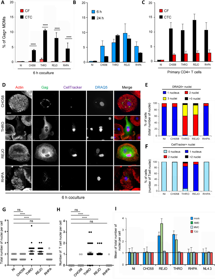
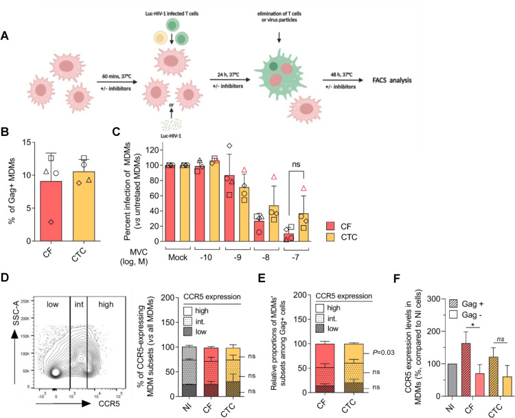
Macrophages (MΦ) are increasingly recognized as HIV-1 target cells involved in the pathogenesis and persistence of infection. Paradoxically, in vitro infection assays suggest that virus isolates are mostly T-cell-tropic and rarely MΦ-tropic. The latter are assumed to emerge under CD4+ T-cell paucity in tissues such as the brain or at late stage when the CD4 T-cell count declines. However, assays to qualify HIV-1 tropism use cell-free viral particles and may not fully reflect the conditions of in vivo MΦ infection through cell-to-cell viral transfer. Here, we investigated the capacity of viruses expressing primary envelope glycoproteins (Envs) with CCR5 and/or CXCR4 usage from different stages of infection, including transmitted/founder Envs, to infect MΦ by a cell-free mode and through cell-to-cell transfer from infected CD4+ T cells. The results show that most viruses were unable to enter MΦ as cell-free particles, in agreement with the current view that non-M-tropic viruses inefficiently use CD4 and/or CCR5 or CXCR4 entry receptors on MΦ. In contrast, all viruses could be effectively cell-to-cell transferred to MΦ from infected CD4+ T cells. We further showed that viral transfer proceeded through Env-dependent cell-cell fusion of infected T cells with MΦ targets, leading to the formation of productively infected multinucleated giant cells. Compared to cell-free infection, infected T-cell/MΦ contacts showed enhanced interactions of R5 M- and non-M-tropic Envs with CD4 and CCR5, resulting in a reduced dependence on receptor expression levels on MΦ for viral entry. Altogether, our results show that virus cell-to-cell transfer overcomes the entry block of isolates initially defined as non-macrophage-tropic, indicating that HIV-1 has a more prevalent tropism for MΦ than initially suggested. This sheds light into the role of this route of virus cell-to-cell transfer to MΦ in CD4+ T cell rich tissues for HIV-1 transmission, dissemination and formation of tissue viral reservoirs.

摘要

巨噬细胞(MΦ)越来越被认为是 HIV-1 的靶细胞,参与感染的发病机制和持续存在。矛盾的是,体外感染实验表明,病毒分离株主要是 T 细胞嗜性的,很少是 MΦ 嗜性的。后者被认为是在组织(如大脑)中 CD4+T 细胞匮乏或 CD4 T 细胞计数下降的晚期出现的。然而,用于确定 HIV-1 嗜性的测定使用无细胞病毒颗粒,并且可能不完全反映通过细胞间病毒转移感染体内 MΦ 的条件。在这里,我们研究了表达不同感染阶段的主要包膜糖蛋白(Env)的病毒(包括传播/原始 Env)通过无细胞模式和从感染的 CD4+T 细胞通过细胞间转移感染 MΦ 的能力。结果表明,大多数病毒无法作为无细胞颗粒进入 MΦ,这与当前的观点一致,即非 M-嗜性病毒在 MΦ 上低效地使用 CD4 和/或 CCR5 或 CXCR4 进入受体。相比之下,所有病毒都可以从感染的 CD4+T 细胞中有效地通过细胞间转移到 MΦ。我们进一步表明,病毒转移是通过感染的 T 细胞与 MΦ 靶细胞之间依赖 Env 的细胞-细胞融合进行的,导致形成具有感染性的多核巨细胞。与无细胞感染相比,感染的 T 细胞/MΦ 接触显示出增强的 R5 M-和非 M-嗜性 Env 与 CD4 和 CCR5 的相互作用,从而减少了对 MΦ 上受体表达水平的依赖性病毒进入。总的来说,我们的结果表明,病毒细胞间转移克服了最初定义为非巨噬细胞嗜性的分离株的进入障碍,表明 HIV-1 对 MΦ 的嗜性比最初建议的更为普遍。这为 HIV-1 在富含 CD4+T 细胞的组织中的传播、扩散和形成组织病毒库的细胞间病毒转移途径的作用提供了线索。

https://cdn.ncbi.nlm.nih.gov/pmc/blobs/b99c/9182568/e5c2457c548c/ppat.1010335.g007.jpg
https://cdn.ncbi.nlm.nih.gov/pmc/blobs/b99c/9182568/7042ae15542b/ppat.1010335.g001.jpg
https://cdn.ncbi.nlm.nih.gov/pmc/blobs/b99c/9182568/534047281228/ppat.1010335.g002.jpg
https://cdn.ncbi.nlm.nih.gov/pmc/blobs/b99c/9182568/9d01acb61c61/ppat.1010335.g003.jpg
https://cdn.ncbi.nlm.nih.gov/pmc/blobs/b99c/9182568/3921dd5d3439/ppat.1010335.g004.jpg
https://cdn.ncbi.nlm.nih.gov/pmc/blobs/b99c/9182568/c11f15dd8048/ppat.1010335.g005.jpg
https://cdn.ncbi.nlm.nih.gov/pmc/blobs/b99c/9182568/f2a9ce6aba39/ppat.1010335.g006.jpg
https://cdn.ncbi.nlm.nih.gov/pmc/blobs/b99c/9182568/e5c2457c548c/ppat.1010335.g007.jpg

相似文献

1
HIV-1 cell-to-cell spread overcomes the virus entry block of non-macrophage-tropic strains in macrophages.HIV-1 细胞间传播可克服非巨噬细胞嗜性株在巨噬细胞中的病毒进入阻断。
PLoS Pathog. 2022 May 27;18(5):e1010335. doi: 10.1371/journal.ppat.1010335. eCollection 2022 May.
2
HIV-1 R5 Macrophage-Tropic Envelope Glycoprotein Trimers Bind CD4 with High Affinity, while the CD4 Binding Site on Non-macrophage-tropic, T-Tropic R5 Envelopes Is Occluded.HIV-1 R5嗜巨噬细胞性包膜糖蛋白三聚体以高亲和力结合CD4,而非巨噬细胞嗜性、T嗜性R5包膜上的CD4结合位点则被封闭。
J Virol. 2018 Jan 2;92(2). doi: 10.1128/JVI.00841-17. Print 2018 Jan 15.
3
Phenotypic Correlates of HIV-1 Macrophage Tropism.HIV-1巨噬细胞嗜性的表型关联
J Virol. 2015 Nov;89(22):11294-311. doi: 10.1128/JVI.00946-15. Epub 2015 Sep 2.
4
Quantification of entry phenotypes of macrophage-tropic HIV-1 across a wide range of CD4 densities.在广泛的CD4密度范围内对嗜巨噬细胞性HIV-1的进入表型进行定量分析。
J Virol. 2014 Feb;88(4):1858-69. doi: 10.1128/JVI.02477-13. Epub 2013 Dec 4.
5
HIV-1 non-macrophage-tropic R5 envelope glycoproteins are not more tropic for entry into primary CD4+ T-cells than envelopes highly adapted for macrophages.与高度适应巨噬细胞的包膜相比,HIV-1非巨噬细胞嗜性R5包膜糖蛋白对进入原代CD4+T细胞的嗜性并不更强。
Retrovirology. 2015 Mar 14;12:25. doi: 10.1186/s12977-015-0141-0.
6
Stoichiometry for entry and binding properties of the Env protein of R5 T cell-tropic HIV-1 and its evolutionary variant of macrophage-tropic HIV-1.包膜蛋白进入和结合性质的化学计量学研究:R5 嗜 T 细胞型 HIV-1 及其进化变体巨噬细胞嗜性 HIV-1。
mBio. 2024 Apr 10;15(4):e0032124. doi: 10.1128/mbio.00321-24. Epub 2024 Mar 1.
7
Tissue-specific sequence alterations in the human immunodeficiency virus type 1 envelope favoring CCR5 usage contribute to persistence of dual-tropic virus in the brain.人类免疫缺陷病毒1型包膜中有利于使用CCR5的组织特异性序列改变有助于双嗜性病毒在大脑中的持续存在。
J Virol. 2009 Jun;83(11):5430-41. doi: 10.1128/JVI.02648-08. Epub 2009 Mar 25.
8
Transmitted/founder and chronic subtype C HIV-1 use CD4 and CCR5 receptors with equal efficiency and are not inhibited by blocking the integrin α4β7.传播/创始者和慢性 C 型 HIV-1 以同等效率使用 CD4 和 CCR5 受体,并且不会被阻断整合素 α4β7 所抑制。
PLoS Pathog. 2012;8(5):e1002686. doi: 10.1371/journal.ppat.1002686. Epub 2012 May 31.
9
Role of CXCR4 in cell-cell fusion and infection of monocyte-derived macrophages by primary human immunodeficiency virus type 1 (HIV-1) strains: two distinct mechanisms of HIV-1 dual tropism.CXCR4在原代人类免疫缺陷病毒1型(HIV-1)毒株介导的单核细胞衍生巨噬细胞的细胞间融合及感染中的作用:HIV-1双嗜性的两种不同机制
J Virol. 1999 Sep;73(9):7117-25. doi: 10.1128/JVI.73.9.7117-7125.1999.
10
Chemokine receptor-usage of clinical HIV-1 isolates obtained from patients with HIV-1 infection in late clinical stages using PHA-blast.使用PHA-母细胞从临床晚期HIV-1感染患者中获得的临床HIV-1分离株的趋化因子受体使用情况
Microbiol Immunol. 1999;43(10):967-74. doi: 10.1111/j.1348-0421.1999.tb03357.x.

引用本文的文献

1
Harnessing miRNA dynamics in HIV-1-infected macrophages: Unveiling new targeted therapeutics using systems biology.利用HIV-1感染巨噬细胞中的微小RNA动态变化:运用系统生物学揭示新的靶向治疗方法。
Comput Struct Biotechnol J. 2025 May 1;27:1754-1771. doi: 10.1016/j.csbj.2025.04.040. eCollection 2025.
2
T cell-mediated SIV dissemination into the CNS: a single-cell transcriptomic analysis.T细胞介导的猴免疫缺陷病毒向中枢神经系统的传播:单细胞转录组分析
Res Sq. 2025 May 9:rs.3.rs-6537361. doi: 10.21203/rs.3.rs-6537361/v1.
3
HIV-1 cell-to-cell infection of macrophages escapes type I interferon and host restriction factors, and is resistant to antiretroviral drugs.

本文引用的文献

1
HIV-1 Vpu Promotes Phagocytosis of Infected CD4 T Cells by Macrophages through Downregulation of CD47.HIV-1 Vpu通过下调CD47促进巨噬细胞对感染的CD4 T细胞的吞噬作用。
mBio. 2021 Aug 31;12(4):e0192021. doi: 10.1128/mBio.01920-21. Epub 2021 Aug 24.
2
The Interplay of HIV-1 and Macrophages in Viral Persistence.HIV-1与巨噬细胞在病毒持续存在中的相互作用
Front Microbiol. 2021 Apr 7;12:646447. doi: 10.3389/fmicb.2021.646447. eCollection 2021.
3
Mechanisms of HIV-1 evasion to the antiviral activity of chemokine CXCL12 indicate potential links with pathogenesis.
HIV-1对巨噬细胞的细胞间感染可逃避I型干扰素和宿主限制因子,并且对抗逆转录病毒药物具有抗性。
PLoS Pathog. 2025 Apr 28;21(4):e1013130. doi: 10.1371/journal.ppat.1013130. eCollection 2025 Apr.
4
CD4+ T cells facilitate replication of primary HIV-1 strains in macrophages and formation of macrophage internal virus-containing compartments.CD4 + T细胞促进原发性HIV-1毒株在巨噬细胞中的复制以及巨噬细胞内病毒包涵体的形成。
J Virol. 2025 Apr 15;99(4):e0018225. doi: 10.1128/jvi.00182-25. Epub 2025 Mar 25.
5
HIV-1 N-myristoylation-dependent hijacking of late endosomes/lysosomes to drive Gag assembly in macrophages.HIV-1 N-豆蔻酰化依赖性晚期内体/溶酶体劫持驱动巨噬细胞中的 Gag 组装。
J Cell Sci. 2024 Nov 15;137(22). doi: 10.1242/jcs.263588. Epub 2024 Nov 21.
6
Characterization of HIV variants from paired Cerebrospinal fluid and Plasma samples in primary microglia and CD4 T-cells.从原代小胶质细胞和 CD4 T 细胞中配对的脑脊液和血浆样本中鉴定 HIV 变异体。
J Neurovirol. 2024 Aug;30(4):380-392. doi: 10.1007/s13365-024-01207-w. Epub 2024 May 7.
7
Macrophages: Key Cellular Players in HIV Infection and Pathogenesis.巨噬细胞:HIV 感染和发病机制中的关键细胞因子。
Viruses. 2024 Feb 13;16(2):288. doi: 10.3390/v16020288.
8
Virus-Induced Cell Fusion and Syncytia Formation.病毒诱导的细胞融合与合胞体形成。
Results Probl Cell Differ. 2024;71:283-318. doi: 10.1007/978-3-031-37936-9_14.
9
Spermine and spermidine bind CXCR4 and inhibit CXCR4- but not CCR5-tropic HIV-1 infection.精胺和亚精胺结合 CXCR4 并抑制 CXCR4-但不抑制 CCR5 嗜性 HIV-1 感染。
Sci Adv. 2023 Jul 7;9(27):eadf8251. doi: 10.1126/sciadv.adf8251. Epub 2023 Jul 5.
10
Productive HIV-1 infection of tissue macrophages by fusion with infected CD4+ T cells.组织巨噬细胞通过与感染的 CD4+T 细胞融合而被 HIV-1 有效感染。
J Cell Biol. 2023 May 1;222(5). doi: 10.1083/jcb.202205103. Epub 2023 Mar 29.
HIV-1 逃避趋化因子 CXCL12 的抗病毒活性的机制表明其与发病机制之间存在潜在联系。
PLoS Pathog. 2021 Apr 19;17(4):e1009526. doi: 10.1371/journal.ppat.1009526. eCollection 2021 Apr.
4
T cell-tropic HIV efficiently infects alveolar macrophages through contact with infected CD4+ T cells.T 细胞嗜性 HIV 通过与感染的 CD4+T 细胞接触,有效地感染肺泡巨噬细胞。
Sci Rep. 2021 Feb 16;11(1):3890. doi: 10.1038/s41598-021-82066-x.
5
Macrophage Cell-Cell Interactions Promoting HIV-1 Infection.巨噬细胞细胞间相互作用促进 HIV-1 感染。
Viruses. 2020 Apr 28;12(5):492. doi: 10.3390/v12050492.
6
A minor population of macrophage-tropic HIV-1 variants is identified in recrudescing viremia following analytic treatment interruption.在分析性治疗中断后出现病毒学反弹时,可鉴定出一小部分巨噬细胞嗜性 HIV-1 变异体。
Proc Natl Acad Sci U S A. 2020 May 5;117(18):9981-9990. doi: 10.1073/pnas.1917034117. Epub 2020 Apr 16.
7
The lipid membrane of HIV-1 stabilizes the viral envelope glycoproteins and modulates their sensitivity to antibody neutralization.HIV-1的脂质膜可稳定病毒包膜糖蛋白,并调节其对抗体中和作用的敏感性。
J Biol Chem. 2020 Jan 10;295(2):348-362. doi: 10.1074/jbc.RA119.009481. Epub 2019 Nov 22.
8
Cell-to-Cell Spreading of HIV-1 in Myeloid Target Cells Escapes SAMHD1 Restriction.HIV-1 在髓样靶细胞中的细胞间传播逃避了 SAMHD1 的限制。
mBio. 2019 Nov 19;10(6):e02457-19. doi: 10.1128/mBio.02457-19.
9
CD4 Incorporation into HIV-1 Viral Particles Exposes Envelope Epitopes Recognized by CD4-Induced Antibodies.CD4 整合入 HIV-1 病毒粒子使包膜表位暴露,这些表位可被 CD4 诱导的抗体识别。
J Virol. 2019 Oct 29;93(22). doi: 10.1128/JVI.01403-19. Print 2019 Nov 15.
10
The HIV Reservoir in Monocytes and Macrophages.HIV 储库:单核细胞和巨噬细胞。
Front Immunol. 2019 Jun 26;10:1435. doi: 10.3389/fimmu.2019.01435. eCollection 2019.