• 文献检索
  • 文档翻译
  • 深度研究
  • 学术资讯
  • Suppr Zotero 插件Zotero 插件
  • 邀请有礼
  • 套餐&价格
  • 历史记录
应用&插件
Suppr Zotero 插件Zotero 插件浏览器插件Mac 客户端Windows 客户端微信小程序
定价
高级版会员购买积分包购买API积分包
服务
文献检索文档翻译深度研究API 文档MCP 服务
关于我们
关于 Suppr公司介绍联系我们用户协议隐私条款
关注我们

Suppr 超能文献

核心技术专利:CN118964589B侵权必究
粤ICP备2023148730 号-1Suppr @ 2026

文献检索

告别复杂PubMed语法,用中文像聊天一样搜索,搜遍4000万医学文献。AI智能推荐,让科研检索更轻松。

立即免费搜索

文件翻译

保留排版,准确专业,支持PDF/Word/PPT等文件格式,支持 12+语言互译。

免费翻译文档

深度研究

AI帮你快速写综述,25分钟生成高质量综述,智能提取关键信息,辅助科研写作。

立即免费体验

蜂巢金标本支持实现对拉长细胞的正交聚焦离子束铣削,以进行冷冻电镜术。

Honeycomb gold specimen supports enabling orthogonal focussed ion beam-milling of elongated cells for cryo-ET.

机构信息

MRC Laboratory of Molecular Biology, Cambridge CB2 0QH, UK.

MRC Laboratory of Molecular Biology, Cambridge CB2 0QH, UK.

出版信息

J Struct Biol. 2024 Jun;216(2):108097. doi: 10.1016/j.jsb.2024.108097. Epub 2024 May 19.

DOI:10.1016/j.jsb.2024.108097
PMID:38772448
原文链接:https://pmc.ncbi.nlm.nih.gov/articles/PMC7616276/
Abstract

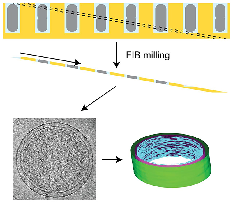
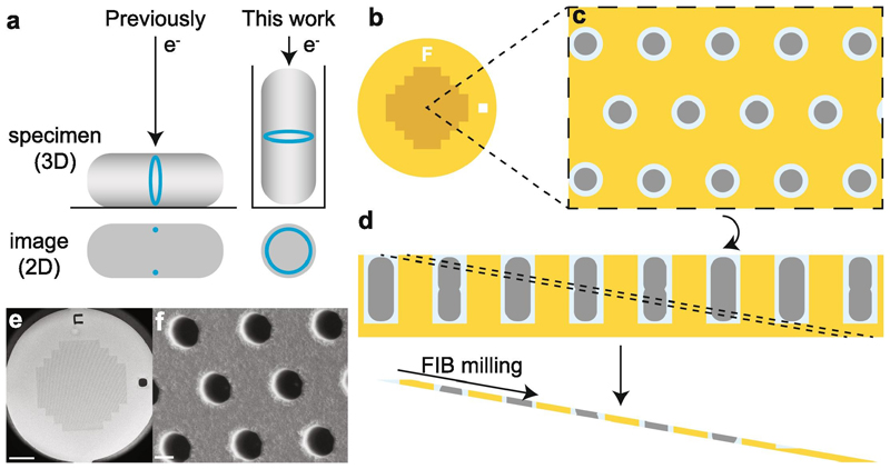
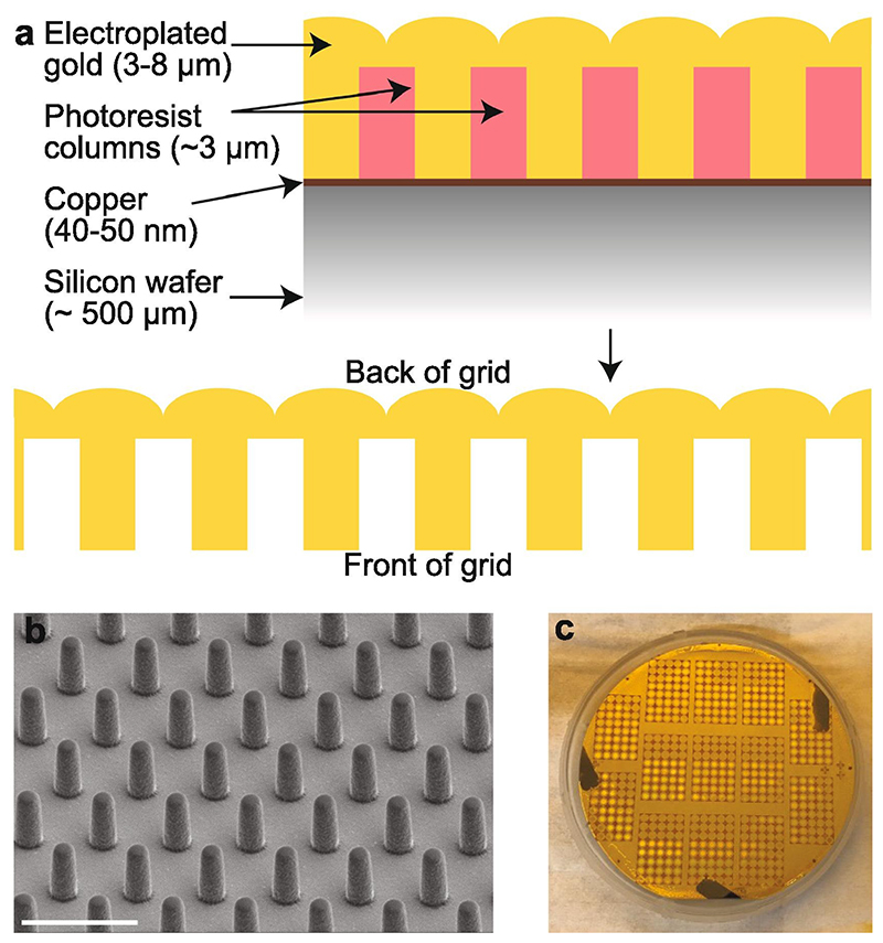
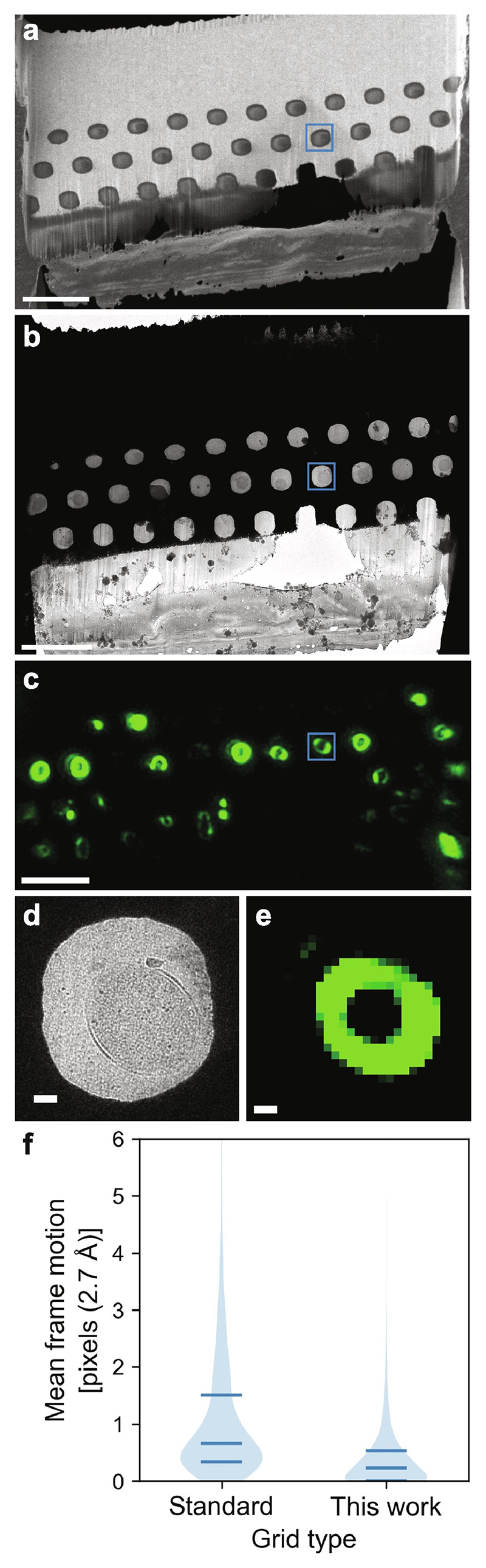
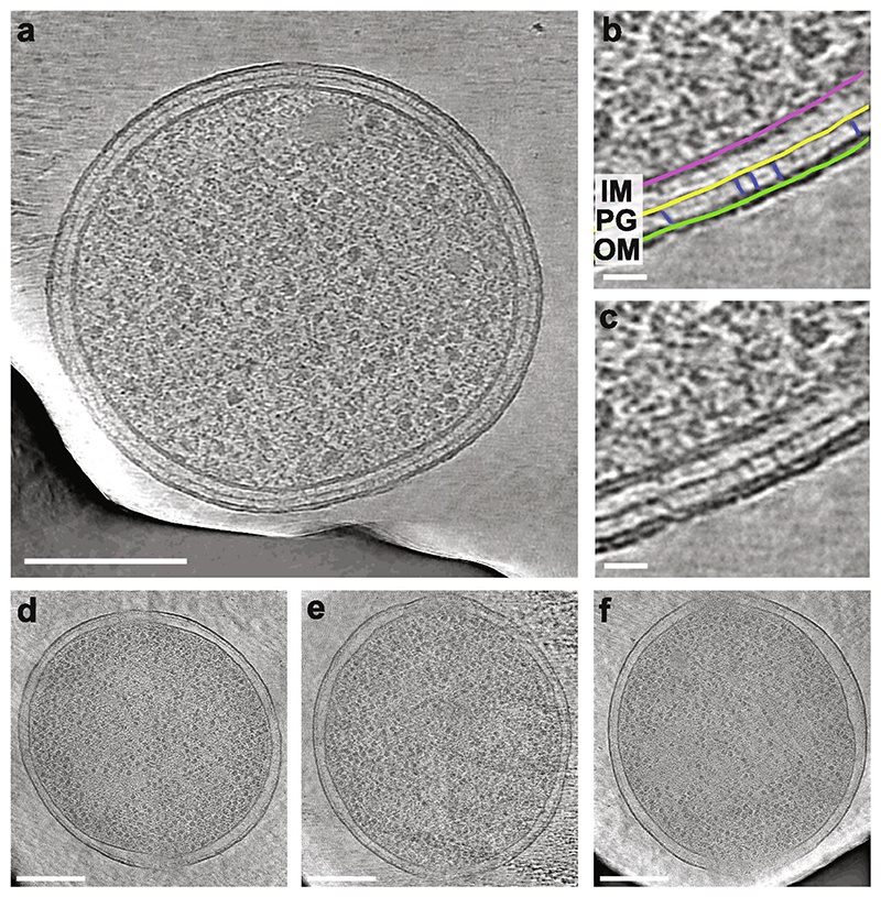
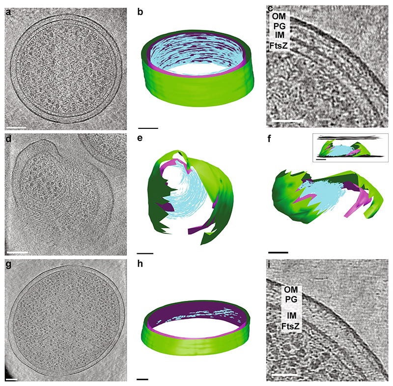
Cryo-focussed ion beam (FIB)-milling is a powerful technique that opens up thick, cellular specimens to high-resolution structural analysis by electron cryotomography (cryo-ET). FIB-milled lamellae can be produced from cells on grids, or cut from thicker, high-pressure frozen specimens. However, these approaches can put geometrical constraints on the specimen that may be unhelpful, particularly when imaging structures within the cell that have a very defined orientation. For example, plunge frozen rod-shaped bacteria orient parallel to the plane of the grid, yet the Z-ring, a filamentous structure of the tubulin-like protein FtsZ and the key organiser of bacterial division, runs around the circumference of the cell such that it is perpendicular to the imaging plane. It is therefore difficult or impractical to image many complete rings with current technologies. To circumvent this problem, we have fabricated monolithic gold specimen supports with a regular array of cylindrical wells in a honeycomb geometry, which trap bacteria in a vertical orientation. These supports, which we call "honeycomb gold discs", replace standard EM grids and when combined with FIB-milling enable the production of lamellae containing cross-sections through cells. The resulting lamellae are more stable and resistant to breakage and charging than conventional lamellae. The design of the honeycomb discs can be modified according to need and so will also enable cryo-ET and cryo-EM imaging of other specimens in otherwise difficult to obtain orientations.

摘要

冷冻聚焦离子束(FIB)-铣削是一种强大的技术,通过电子冷冻断层扫描(cryo-ET)可以将厚的、细胞状的样本开放到高分辨率的结构分析中。FIB 铣削的薄片可以从网格上的细胞中产生,也可以从较厚的、高压冷冻的样本中切割。然而,这些方法可能会对样本施加几何约束,这可能是无益的,特别是在对具有非常确定取向的细胞内结构进行成像时。例如, plunge 冷冻的棒状细菌与网格平面平行取向,然而,Z 环,一种类似于微管蛋白的 FtsZ 纤维状结构,是细菌分裂的关键组织者,它环绕细胞的周长延伸,因此与成像平面垂直。因此,用当前的技术很难或不可能对许多完整的环进行成像。为了克服这个问题,我们制造了具有蜂窝状几何形状的规则圆柱形孔阵列的整体金制样本支架,将细菌固定在垂直方向。这些支架,我们称之为“蜂窝金盘”,替代了标准的 EM 网格,并且与 FIB 铣削结合使用,可以生产出包含细胞横截面的薄片。所得的薄片比传统的薄片更稳定,不易断裂和充电。蜂窝盘的设计可以根据需要进行修改,因此也将能够对其他在难以获得的取向中进行 cryo-ET 和 cryo-EM 成像。

https://cdn.ncbi.nlm.nih.gov/pmc/blobs/2bf9/7616276/97dc298f14cf/EMS197563-f006.jpg
https://cdn.ncbi.nlm.nih.gov/pmc/blobs/2bf9/7616276/5cc2c1dd6f55/EMS197563-f007.jpg
https://cdn.ncbi.nlm.nih.gov/pmc/blobs/2bf9/7616276/05033aeb5cd3/EMS197563-f001.jpg
https://cdn.ncbi.nlm.nih.gov/pmc/blobs/2bf9/7616276/68c69b753f6c/EMS197563-f002.jpg
https://cdn.ncbi.nlm.nih.gov/pmc/blobs/2bf9/7616276/b0a54d78db5c/EMS197563-f003.jpg
https://cdn.ncbi.nlm.nih.gov/pmc/blobs/2bf9/7616276/cbe899cd29b3/EMS197563-f004.jpg
https://cdn.ncbi.nlm.nih.gov/pmc/blobs/2bf9/7616276/a810f58517e5/EMS197563-f005.jpg
https://cdn.ncbi.nlm.nih.gov/pmc/blobs/2bf9/7616276/97dc298f14cf/EMS197563-f006.jpg

相似文献

1
Honeycomb gold specimen supports enabling orthogonal focussed ion beam-milling of elongated cells for cryo-ET.蜂巢金标本支持实现对拉长细胞的正交聚焦离子束铣削,以进行冷冻电镜术。
J Struct Biol. 2024 Jun;216(2):108097. doi: 10.1016/j.jsb.2024.108097. Epub 2024 May 19.
2
Robust workflow and instrumentation for cryo-focused ion beam milling of samples for electron cryotomography.用于电子冷冻断层扫描样品的冷冻聚焦离子束铣削的强大工作流程和仪器设备。
Ultramicroscopy. 2018 Jul;190:1-11. doi: 10.1016/j.ultramic.2018.04.002. Epub 2018 Apr 6.
3
Mind the gap: Micro-expansion joints drastically decrease the bending of FIB-milled cryo-lamellae.注意间隙:微膨胀节可大大减少 FIB 铣削的低温薄片的弯曲。
J Struct Biol. 2019 Dec 1;208(3):107389. doi: 10.1016/j.jsb.2019.09.006. Epub 2019 Sep 16.
4
Cryo-FIB specimen preparation for use in a cartridge-type cryo-TEM.用于盒式冷冻透射电子显微镜的冷冻聚焦离子束标本制备
J Struct Biol. 2017 Aug;199(2):114-119. doi: 10.1016/j.jsb.2017.05.011. Epub 2017 May 27.
5
Sample Preparation by 3D-Correlative Focused Ion Beam Milling for High-Resolution Cryo-Electron Tomography.通过 3D 相关聚焦离子束铣削进行高分辨率冷冻电子断层成像的样品制备。
J Vis Exp. 2021 Oct 25(176). doi: 10.3791/62886.
6
Quantification of gallium cryo-FIB milling damage in biological lamellae.定量分析生物切片中镓冷冻聚焦离子束铣削损伤。
Proc Natl Acad Sci U S A. 2023 Jun 6;120(23):e2301852120. doi: 10.1073/pnas.2301852120. Epub 2023 May 22.
7
Ultrastructure of human brain tissue vitrified from autopsy revealed by cryo-ET with cryo-plasma FIB milling.利用冷冻等离子体 FIB 铣削的 cryo-ET 技术揭示了从尸检中玻璃化的人类脑组织的超微结构。
Nat Commun. 2024 Mar 26;15(1):2660. doi: 10.1038/s41467-024-47066-1.
8
Automated cryo-lamella preparation for high-throughput in-situ structural biology.自动化低温薄片制备用于高通量原位结构生物学。
J Struct Biol. 2020 May 1;210(2):107488. doi: 10.1016/j.jsb.2020.107488. Epub 2020 Feb 29.
9
Sample preparation and image registration for correlative cryo-FM and cryo-FIB-SEM of plunge-frozen mammalian cells.用于哺乳动物细胞冷冻深剖样品的冷冻-FM 和冷冻-FIB-SEM 相关的样品制备和图像配准。
STAR Protoc. 2022 Feb 8;3(1):101142. doi: 10.1016/j.xpro.2022.101142. eCollection 2022 Mar 18.
10
Waffle Method: A general and flexible approach for improving throughput in FIB-milling.华夫饼法:提高 FIB 铣削效率的通用且灵活的方法。
Nat Commun. 2022 Apr 6;13(1):1857. doi: 10.1038/s41467-022-29501-3.

本文引用的文献

1
Serial Lift-Out: sampling the molecular anatomy of whole organisms.连续提取:对整个生物体的分子解剖结构进行采样。
Nat Methods. 2024 Sep;21(9):1684-1692. doi: 10.1038/s41592-023-02113-5. Epub 2023 Dec 18.
2
The reduction of FIB damage on cryo-lamella by lowering energy of ion beam revealed by a quantitative analysis.通过定量分析揭示了降低离子束能量可减少冷冻薄片上的FIB损伤。
Structure. 2023 Oct 5;31(10):1275-1281.e4. doi: 10.1016/j.str.2023.07.002. Epub 2023 Jul 31.
3
Quantification of gallium cryo-FIB milling damage in biological lamellae.
定量分析生物切片中镓冷冻聚焦离子束铣削损伤。
Proc Natl Acad Sci U S A. 2023 Jun 6;120(23):e2301852120. doi: 10.1073/pnas.2301852120. Epub 2023 May 22.
4
OpenFIBSEM: A universal API for FIBSEM control.OpenFIBSEM:用于 FIBSEM 控制的通用 API。
J Struct Biol. 2023 Sep;215(3):107967. doi: 10.1016/j.jsb.2023.107967. Epub 2023 Apr 25.
5
Plasma FIB milling for the determination of structures in situ.等离子体 FIB 铣削用于原位结构的测定。
Nat Commun. 2023 Feb 6;14(1):629. doi: 10.1038/s41467-023-36372-9.
6
ELI trifocal microscope: a precise system to prepare target cryo-lamellae for in situ cryo-ET study.ELI 三焦点显微镜:用于原位冷冻电镜研究的目标冷冻薄片制备的精确系统。
Nat Methods. 2023 Feb;20(2):276-283. doi: 10.1038/s41592-022-01748-0. Epub 2023 Jan 16.
7
In situ cryo-FIB/SEM Specimen Preparation Using the Waffle Method.使用华夫饼法进行原位冷冻聚焦离子束/扫描电子显微镜标本制备。
Bio Protoc. 2022 Nov 5;12(21). doi: 10.21769/BioProtoc.4544.
8
A Bayesian approach to single-particle electron cryo-tomography in RELION-4.0.基于 RELION-4.0 的单颗粒电子冷冻断层成像的贝叶斯方法。
Elife. 2022 Dec 5;11:e83724. doi: 10.7554/eLife.83724.
9
Structures of the eukaryotic ribosome and its translational states in situ.真核核糖体及其原位翻译状态的结构。
Nat Commun. 2022 Dec 2;13(1):7435. doi: 10.1038/s41467-022-34997-w.
10
Parallel cryo electron tomography on in situ lamellae.原位薄片的平行冷冻电子断层扫描术
Nat Methods. 2023 Jan;20(1):131-138. doi: 10.1038/s41592-022-01690-1. Epub 2022 Dec 1.