Department of Radiological Sciences, International University of Health and Welfare, 2600-1 Kitakanemaru, Otawara, Tochigi, 324-8501, Japan.
Center of Medical Ultrasonics, Dokkyo Medical University, Mibu, Tochigi, Japan.
J Med Ultrason (2001). 2024 Oct;51(4):567-580. doi: 10.1007/s10396-024-01443-x. Epub 2024 May 23.
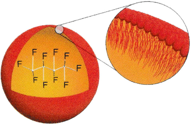
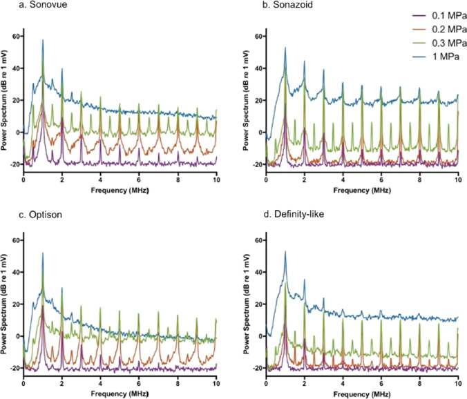
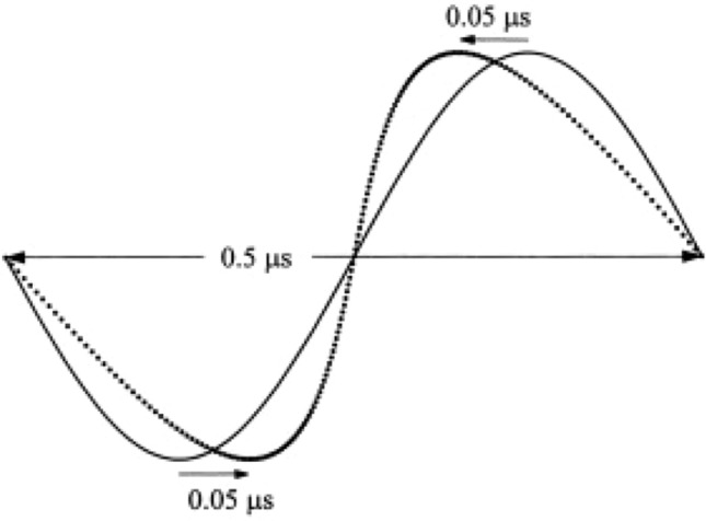
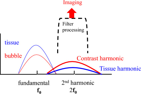
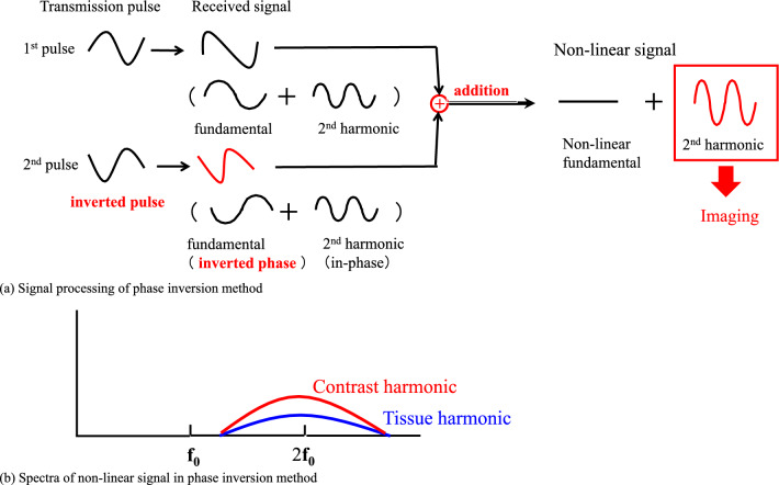
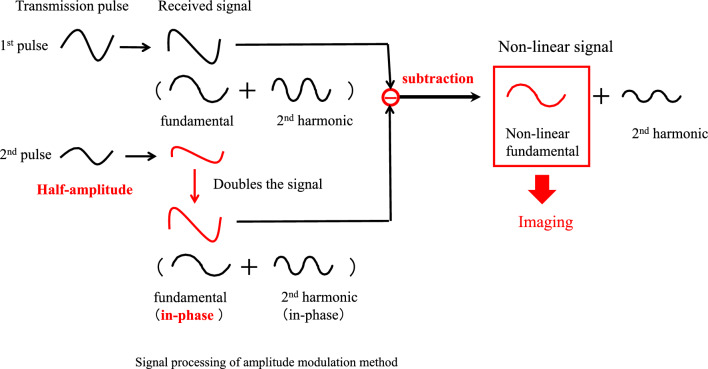
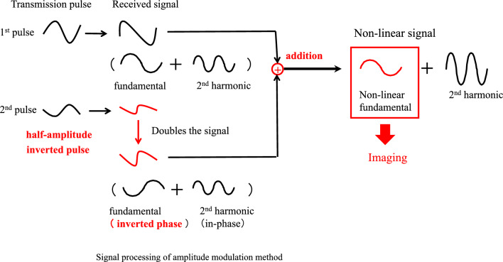
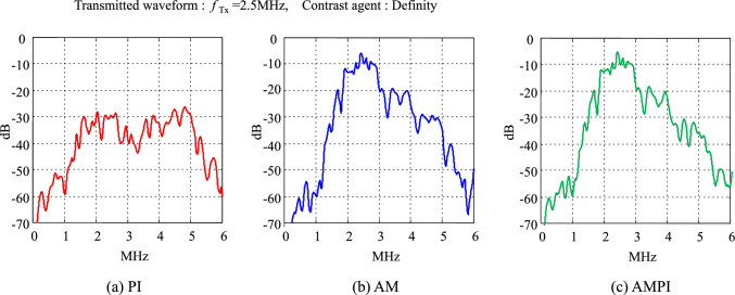
Sonazoid, an ultrasound contrast agent, has been covered by insurance in Japan since January 2007 for the diagnosis of hepatic mass lesions and is widely used for diagnosing not only primary liver cancer but also liver metastases such as those from breast cancer and colorectal cancer. Contrast-enhanced ultrasound for breast mass lesions has been covered by insurance since August 2012 after phase II and phase III clinical trials showed that the diagnostic performance was significantly superior to that of B-mode and contrast-enhanced magnetic resonance imaging. This paper describes the principles of imaging techniques in contrast-enhanced ultrasonography including the filter, pulse inversion, amplitude modulation, and amplitude-modulated pulse inversion methods. The pulse inversion method, which visualizes the second-harmonic component using the nonlinear scattering characteristics of the contrast agent, is widely used regardless of the contrast agent and target organ because of its high resolution. Sonazoid has a stiffer shell and requires a higher acoustic amplitude than Sonovue to generate nonlinear vibrations. The higher transmitted sound pressure generates more tissue harmonic components. Since pulse inversion allows visualization of the tissue harmonic components, amplitude modulation and amplitude-modulated pulse inversion, which include few tissue harmonic components, are primarily used. Amplitude modulation methods detect nonlinear signals from the contrast agent in the fundamental band. The mechanism of the amplitude modulation is considered to be changes in the echo signal's phase depending on the sound pressure. Since the tissue-derived component is minor in amplitude modulation methods, good contrast sensitivity can be obtained.
声诺维,一种超声造影剂,自 2007 年 1 月起在日本被纳入医保,用于诊断肝脏肿块病变,已被广泛用于诊断原发性肝癌,以及乳腺癌和结直肠癌等肝转移瘤。增强超声用于乳腺肿块病变的诊断自 2012 年 8 月起被纳入医保,此前 II 期和 III 期临床试验表明,该诊断性能明显优于 B 型超声和对比增强磁共振成像。本文介绍了增强超声成像技术的原理,包括滤波、脉冲反相、调幅和调幅脉冲反相方法。脉冲反相法利用造影剂的非线性散射特性来可视化二次谐波分量,无论造影剂和靶器官如何,由于其具有高分辨率,因此被广泛应用。声诺维具有更硬的外壳,与声诺维相比,需要更高的声幅来产生非线性振动。更高的声压产生更多的组织谐波分量。由于脉冲反相允许可视化组织谐波分量,因此主要使用调幅和调幅脉冲反相,它们包含很少的组织谐波分量。调幅方法检测基频带中造影剂的非线性信号。调幅的机制被认为是根据声压改变回波信号的相位。由于调幅方法中的组织分量幅度较小,因此可以获得良好的对比敏感度。