Salvante Enrica Raffaella Grazia, Popoiu Anca Voichita, Saxena Amulya K, Popoiu Tudor Alexandru, Boia Eugen Sorin, Cimpean Anca Maria, Rus Florina Stefania, Dorobantu Florica Ramona, Chis Monica
Doctoral School, Victor Babes University of Medicine and Pharmacy Timisoara, 300041 Timisoara, Romania.
Emergency Hospital for Children Louis Turcanu, 300011 Timisoara, Romania.
Bioengineering (Basel). 2024 Apr 25;11(5):423. doi: 10.3390/bioengineering11050423.
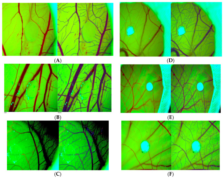
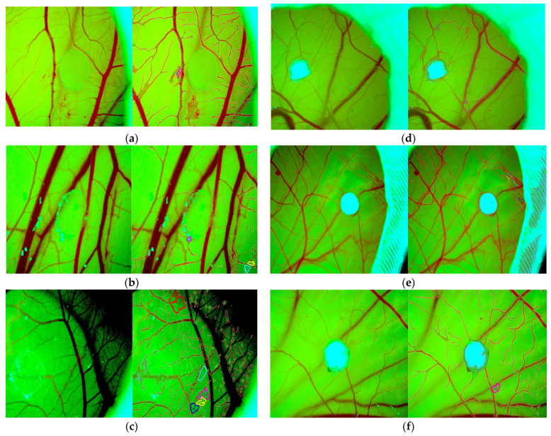
Type I collagen, prevalent in the extracellular matrix, is biocompatible and crucial for tissue engineering and wound healing, including angiogenesis and vascular maturation/stabilization as required processes of newly formed tissue constructs or regeneration. Sometimes, improper vascularization causes unexpected outcomes. Vascularization failure may be caused by extracellular matrix collagen and non-collagen components heterogeneously. This study compares the angiogenic potential of collagen type I-based scaffolds and collagen type I/glycosaminoglycans scaffolds by using the chick embryo chorioallantoic membrane (CAM) model and IKOSA digital image analysis. Two clinically used biomaterials, Xenoderm (containing type I collagen derived from decellularized porcine extracellular matrix) and a dual-layer collagen sponge (DLC, with a biphasic composition of type I collagen combined with glycosaminoglycans) were tested for their ability to induce new vascular network formation. The AI-based IKOSA app enhanced the research by calculating from stereomicroscopic images angiogenic parameters such as total vascular area, branching sites, vessel length, and vascular thickness. The study confirmed that Xenoderm caused a fast angiogenic response and substantial vascular growth, but was unable to mature the vascular network. DLC scaffold, in turn, produced a slower angiogenic response, but a more steady and organic vascular maturation and stabilization. This research can improve collagen-based knowledge by better assessing angiogenesis processes. DLC may be preferable to Xenoderm or other materials for functional neovascularization, according to the findings.
I型胶原蛋白在细胞外基质中普遍存在,具有生物相容性,对组织工程和伤口愈合至关重要,包括血管生成以及血管成熟/稳定,这些都是新形成的组织构建物或再生所需的过程。有时,血管化不当会导致意外结果。血管化失败可能是由细胞外基质中的胶原蛋白和非胶原蛋白成分不均匀引起的。本研究通过使用鸡胚绒毛尿囊膜(CAM)模型和IKOSA数字图像分析,比较了I型胶原蛋白基支架和I型胶原蛋白/糖胺聚糖支架的血管生成潜力。测试了两种临床使用的生物材料,即Xenoderm(含有源自脱细胞猪细胞外基质的I型胶原蛋白)和双层胶原海绵(DLC,具有I型胶原蛋白与糖胺聚糖的双相组成)诱导新血管网络形成的能力。基于人工智能的IKOSA应用程序通过从立体显微镜图像计算血管生成参数,如总血管面积、分支点、血管长度和血管厚度,增强了研究。该研究证实,Xenoderm引起快速的血管生成反应和大量的血管生长,但无法使血管网络成熟。相反,DLC支架产生较慢的血管生成反应,但血管成熟和稳定更为稳定和有机。这项研究可以通过更好地评估血管生成过程来增进对胶原蛋白的了解。根据研究结果,在功能性新血管形成方面,DLC可能比Xenoderm或其他材料更具优势。