• 文献检索
  • 文档翻译
  • 深度研究
  • 学术资讯
  • Suppr Zotero 插件Zotero 插件
  • 邀请有礼
  • 套餐&价格
  • 历史记录
应用&插件
Suppr Zotero 插件Zotero 插件浏览器插件Mac 客户端Windows 客户端微信小程序
定价
高级版会员购买积分包购买API积分包
服务
文献检索文档翻译深度研究API 文档MCP 服务
关于我们
关于 Suppr公司介绍联系我们用户协议隐私条款
关注我们

Suppr 超能文献

核心技术专利:CN118964589B侵权必究
粤ICP备2023148730 号-1Suppr @ 2026

文献检索

告别复杂PubMed语法,用中文像聊天一样搜索,搜遍4000万医学文献。AI智能推荐,让科研检索更轻松。

立即免费搜索

文件翻译

保留排版,准确专业,支持PDF/Word/PPT等文件格式,支持 12+语言互译。

免费翻译文档

深度研究

AI帮你快速写综述,25分钟生成高质量综述,智能提取关键信息,辅助科研写作。

立即免费体验

HIV-1 衣壳蛋白迅速诱导非分裂细胞中长寿命 CPSF6 斑点,但在未感染的 T 细胞中已经存在类似的斑点。

HIV-1 Capsid Rapidly Induces Long-Lived CPSF6 Puncta in Non-Dividing Cells, but Similar Puncta Already Exist in Uninfected T-Cells.

机构信息

Retroviral Replication Laboratory, The Francis Crick Institute, London NW1 1AT, UK.

出版信息

Viruses. 2024 Apr 25;16(5):670. doi: 10.3390/v16050670.

DOI:10.3390/v16050670
PMID:38793552
原文链接:https://pmc.ncbi.nlm.nih.gov/articles/PMC11125723/
Abstract

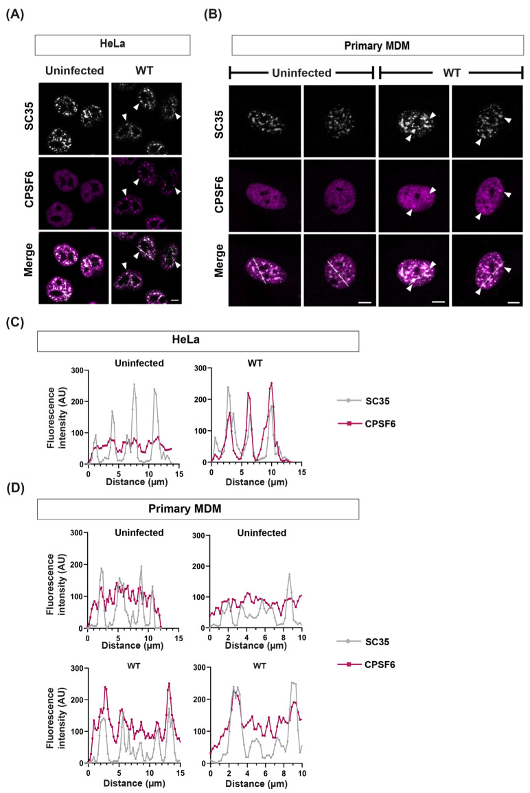
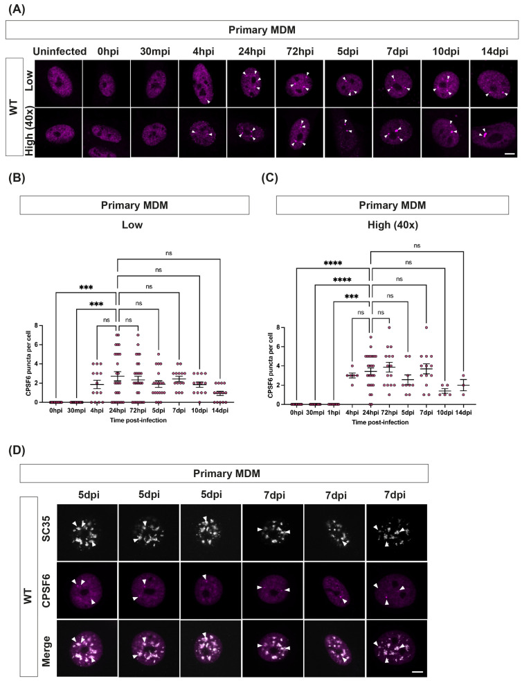
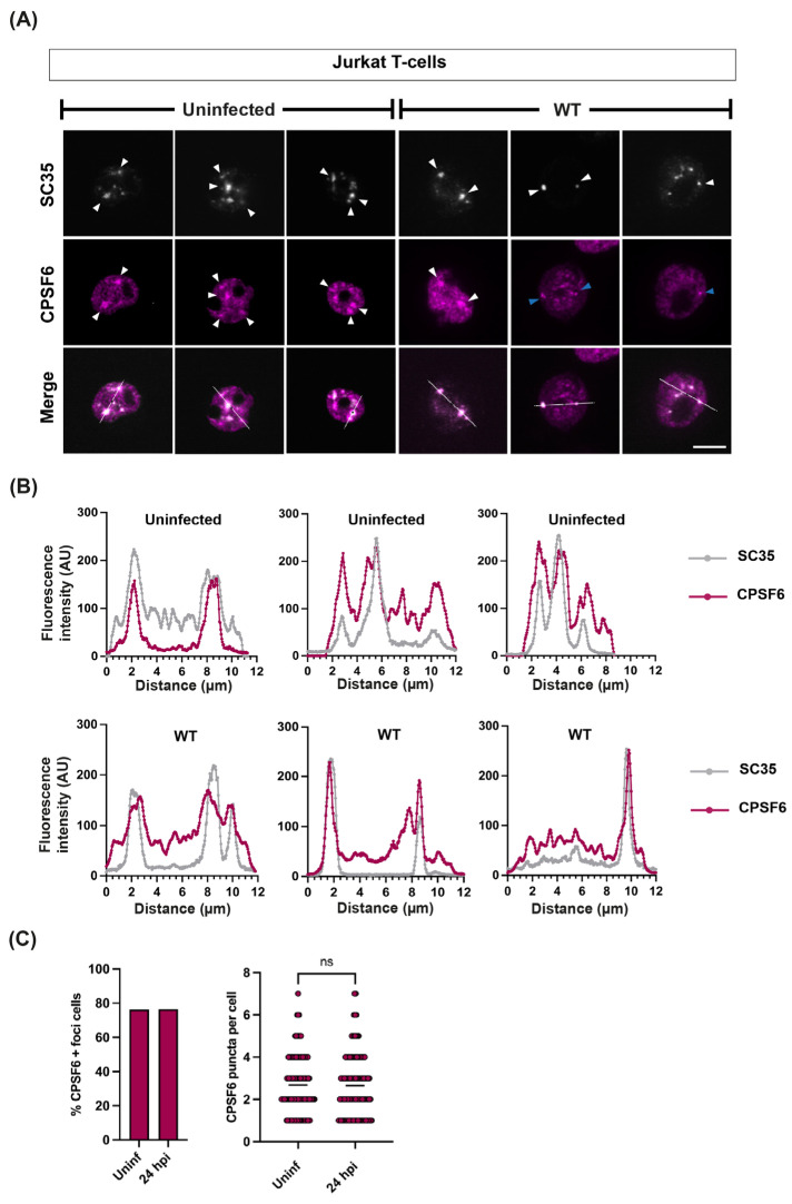
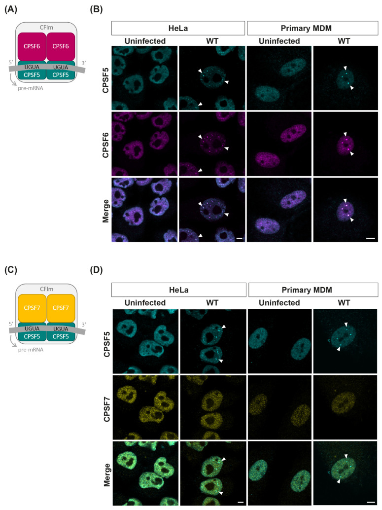
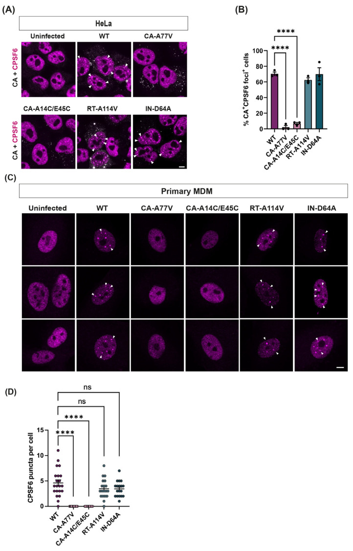
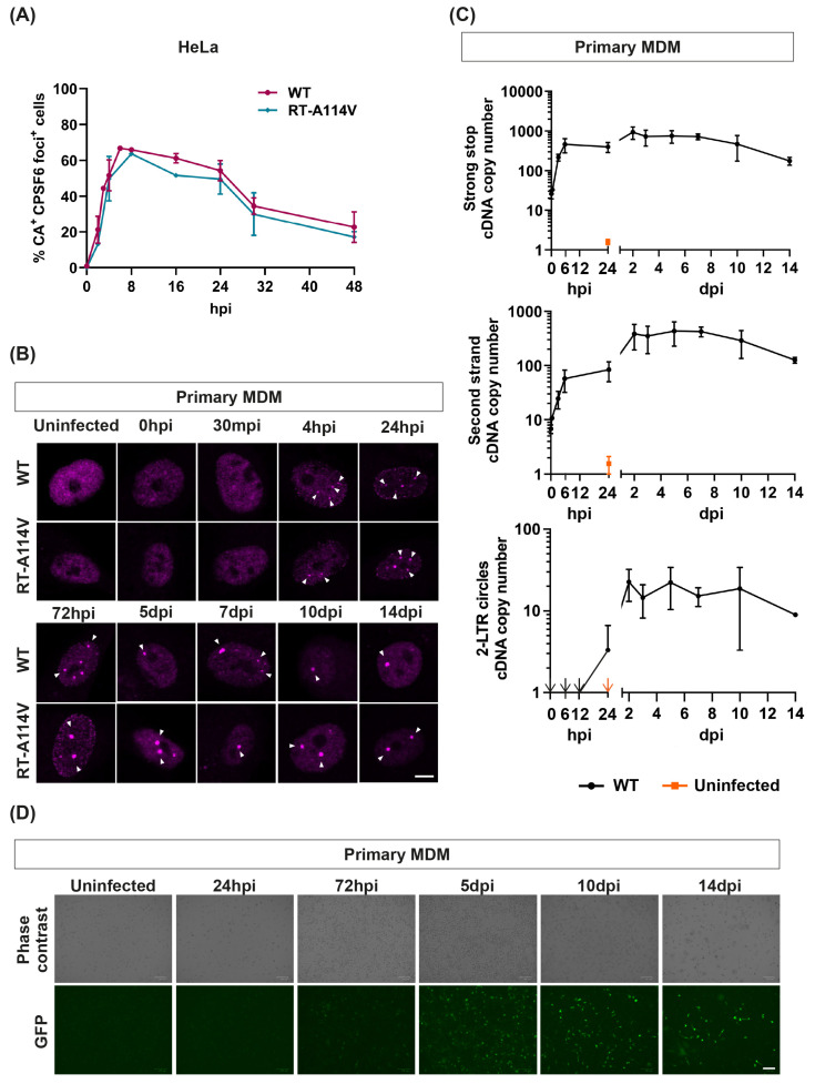
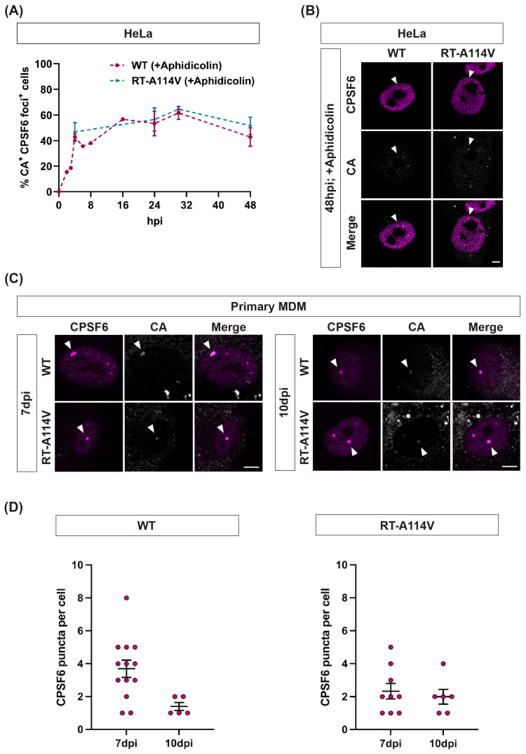
The HIV-1 capsid (CA) protein forms the outer shell of the viral core that is released into the cytoplasm upon infection. CA binds various cellular proteins, including CPSF6, that direct HIV-1 integration into speckle-associated domains in host chromatin. Upon HIV-1 infection, CPSF6 forms puncta in the nucleus. Here, we characterised these CPSF6 puncta further in HeLa cells, T-cells and macrophages and confirmed that integration and reverse transcription are not required for puncta formation. Indeed, we found that puncta formed very rapidly after infection, correlating with the time that CA entered the nucleus. In aphidicolin-treated HeLa cells and macrophages, puncta were detected for the length of the experiment, suggesting that puncta are only lost upon cell division. CA still co-localised with CPSF6 puncta at the latest time points, considerably after the peak of reverse transcription and integration. Intriguingly, the number of puncta induced in macrophages did not correlate with the MOI or the total number of nuclear speckles present in each cell, suggesting that CA/CPSF6 is only directed to a few nuclear speckles. Furthermore, we found that CPSF6 already co-localised with nuclear speckles in uninfected T-cells, suggesting that HIV-1 promotes a natural behaviour of CPSF6.

摘要

HIV-1 衣壳(CA)蛋白形成病毒核心的外壳,在感染时释放到细胞质中。CA 结合各种细胞蛋白,包括 CPSF6,指导 HIV-1 整合到宿主染色质中的斑点相关结构域中。在 HIV-1 感染后,CPSF6 在核内形成斑点。在这里,我们进一步在 HeLa 细胞、T 细胞和巨噬细胞中对这些 CPSF6 斑点进行了表征,并证实整合和逆转录不是形成斑点所必需的。事实上,我们发现斑点在感染后很快形成,与 CA 进入核内的时间相关。在用阿非迪可林处理的 HeLa 细胞和巨噬细胞中,斑点在实验过程中一直存在,表明只有在细胞分裂时才会丢失斑点。CA 仍然与 CPSF6 斑点共定位,时间远远晚于逆转录和整合的高峰期。有趣的是,巨噬细胞中诱导的斑点数量与 MOI 或每个细胞中存在的核斑点总数无关,这表明 CA/CPSF6 仅被引导到少数核斑点。此外,我们发现未感染的 T 细胞中 CPSF6 已经与核斑点共定位,这表明 HIV-1 促进了 CPSF6 的自然行为。

https://cdn.ncbi.nlm.nih.gov/pmc/blobs/e4e3/11125723/ccb1e6d1eda4/viruses-16-00670-g007.jpg
https://cdn.ncbi.nlm.nih.gov/pmc/blobs/e4e3/11125723/a3a0a8033ec9/viruses-16-00670-g001.jpg
https://cdn.ncbi.nlm.nih.gov/pmc/blobs/e4e3/11125723/2fbc9d610d7e/viruses-16-00670-g002.jpg
https://cdn.ncbi.nlm.nih.gov/pmc/blobs/e4e3/11125723/d2610ef9cdfc/viruses-16-00670-g003.jpg
https://cdn.ncbi.nlm.nih.gov/pmc/blobs/e4e3/11125723/6e46b06a8203/viruses-16-00670-g004.jpg
https://cdn.ncbi.nlm.nih.gov/pmc/blobs/e4e3/11125723/2fcf97121c48/viruses-16-00670-g005.jpg
https://cdn.ncbi.nlm.nih.gov/pmc/blobs/e4e3/11125723/7a0af0223e3d/viruses-16-00670-g006.jpg
https://cdn.ncbi.nlm.nih.gov/pmc/blobs/e4e3/11125723/ccb1e6d1eda4/viruses-16-00670-g007.jpg

相似文献

1
HIV-1 Capsid Rapidly Induces Long-Lived CPSF6 Puncta in Non-Dividing Cells, but Similar Puncta Already Exist in Uninfected T-Cells.HIV-1 衣壳蛋白迅速诱导非分裂细胞中长寿命 CPSF6 斑点,但在未感染的 T 细胞中已经存在类似的斑点。
Viruses. 2024 Apr 25;16(5):670. doi: 10.3390/v16050670.
2
Formation of nuclear CPSF6/CPSF5 biomolecular condensates upon HIV-1 entry into the nucleus is important for productive infection.核 CPSF6/CPSF5 生物分子凝聚物的形成对于 HIV-1 进入细胞核后的有效感染很重要。
Sci Rep. 2023 Jul 6;13(1):10974. doi: 10.1038/s41598-023-37364-x.
3
HIV-1 usurps mixed-charge domain-dependent CPSF6 phase separation for higher-order capsid binding, nuclear entry and viral DNA integration.HIV-1 劫持混合电荷域依赖性 CPSF6 相分离以增强衣壳结合、核进入和病毒 DNA 整合。
Nucleic Acids Res. 2024 Oct 14;52(18):11060-11082. doi: 10.1093/nar/gkae769.
4
Truncated CPSF6 Forms Higher-Order Complexes That Bind and Disrupt HIV-1 Capsid.截短的 CPSF6 形成更高级的复合物,结合并破坏 HIV-1 衣壳。
J Virol. 2018 Jun 13;92(13). doi: 10.1128/JVI.00368-18. Print 2018 Jul 1.
5
Capsid-CPSF6 Interaction Is Dispensable for HIV-1 Replication in Primary Cells but Is Selected during Virus Passage In Vivo.衣壳与CPSF6的相互作用对HIV-1在原代细胞中的复制并非必需,但在体内病毒传代过程中会被选择。
J Virol. 2016 Jul 11;90(15):6918-6935. doi: 10.1128/JVI.00019-16. Print 2016 Aug 1.
6
Cytoplasmic CPSF6 Regulates HIV-1 Capsid Trafficking and Infection in a Cyclophilin A-Dependent Manner.细胞质 CPSF6 通过依赖环孢素 A 的方式调节 HIV-1 衣壳的运输和感染。
mBio. 2021 Mar 23;12(2):e03142-20. doi: 10.1128/mBio.03142-20.
7
The Cleavage and Polyadenylation Specificity Factor 6 (CPSF6) Subunit of the Capsid-recruited Pre-messenger RNA Cleavage Factor I (CFIm) Complex Mediates HIV-1 Integration into Genes.衣壳招募的信使前体RNA切割因子I(CFIm)复合物的切割和聚腺苷酸化特异性因子6(CPSF6)亚基介导HIV-1整合入基因。
J Biol Chem. 2016 May 27;291(22):11809-19. doi: 10.1074/jbc.M116.721647. Epub 2016 Mar 18.
8
Analysis of CA Content and CPSF6 Dependence of Early HIV-1 Replication Complexes in SupT1-R5 Cells.分析 SupT1-R5 细胞中早期 HIV-1 复制复合物的 CA 含量和 CPSF6 依赖性。
mBio. 2019 Nov 5;10(6):e02501-19. doi: 10.1128/mBio.02501-19.
9
Detection of CPSF6 in Biomolecular Condensates as a Reporter of HIV-1 Nuclear Import.检测生物分子凝聚物中的 CPSF6 作为 HIV-1 核输入的报告分子。
Methods Mol Biol. 2024;2807:127-138. doi: 10.1007/978-1-0716-3862-0_9.
10
HIV-1 nuclear import in macrophages is regulated by CPSF6-capsid interactions at the nuclear pore complex.HIV-1 核输入受核孔复合物中 CPSF6-衣壳相互作用的调节。
Elife. 2019 Jan 23;8:e41800. doi: 10.7554/eLife.41800.

引用本文的文献

1
HIV-1 Infection Regulates Gene Expression by Altering Alternative Polyadenylation Through CPSF6 and CPSF5 Delocalization.HIV-1感染通过CPSF6和CPSF5的重新定位改变可变聚腺苷酸化来调节基因表达。
bioRxiv. 2025 Aug 7:2025.08.07.669137. doi: 10.1101/2025.08.07.669137.
2
Retrointegration2023-Papers from the 7th International Conference on Retroviral Integration.《逆转录病毒整合2023——第七届逆转录病毒整合国际会议论文集》
Viruses. 2025 Jun 23;17(7):879. doi: 10.3390/v17070879.
3
The role of chromatin in retroviral preintegration complex function.

本文引用的文献

1
Genome organization around nuclear speckles drives mRNA splicing efficiency.基因组在核斑周围的组织驱动 mRNA 剪接效率。
Nature. 2024 May;629(8014):1165-1173. doi: 10.1038/s41586-024-07429-6. Epub 2024 May 8.
2
Emerging roles of nuclear bodies in genome spatial organization.核体在基因组空间组织中的新兴作用。
Trends Cell Biol. 2024 Jul;34(7):595-605. doi: 10.1016/j.tcb.2023.10.012. Epub 2023 Nov 21.
3
CPSF6 regulates alternative polyadenylation and proliferation of cancer cells through phase separation.CPSF6 通过相分离调节癌细胞的可变多聚腺苷酸化和增殖。
染色质在逆转录病毒整合前复合体功能中的作用。
J Biol Chem. 2025 Jul 3;301(8):110440. doi: 10.1016/j.jbc.2025.110440.
4
Disruption of CPSF6 enhances cellular permissivity to HIV-1 infection through alternative polyadenylation.CPSF6的破坏通过可变多聚腺苷酸化增强细胞对HIV-1感染的易感性。
Res Sq. 2024 Dec 4:rs.3.rs-5099896. doi: 10.21203/rs.3.rs-5099896/v1.
5
HIV-1 usurps mixed-charge domain-dependent CPSF6 phase separation for higher-order capsid binding, nuclear entry and viral DNA integration.HIV-1 劫持混合电荷域依赖性 CPSF6 相分离以增强衣壳结合、核进入和病毒 DNA 整合。
Nucleic Acids Res. 2024 Oct 14;52(18):11060-11082. doi: 10.1093/nar/gkae769.
Cell Rep. 2023 Oct 31;42(10):113197. doi: 10.1016/j.celrep.2023.113197. Epub 2023 Sep 30.
4
Formation of nuclear CPSF6/CPSF5 biomolecular condensates upon HIV-1 entry into the nucleus is important for productive infection.核 CPSF6/CPSF5 生物分子凝聚物的形成对于 HIV-1 进入细胞核后的有效感染很重要。
Sci Rep. 2023 Jul 6;13(1):10974. doi: 10.1038/s41598-023-37364-x.
5
HIV-Induced CPSF6 Condensates.HIV 诱导的 CPSF6 凝聚物。
J Mol Biol. 2023 Aug 15;435(16):168094. doi: 10.1016/j.jmb.2023.168094. Epub 2023 Apr 14.
6
The capsid lattice engages a bipartite NUP153 motif to mediate nuclear entry of HIV-1 cores.衣壳晶格与二聚体 NUP153 基序结合,介导 HIV-1 核心进入细胞核。
Proc Natl Acad Sci U S A. 2023 Mar 28;120(13):e2202815120. doi: 10.1073/pnas.2202815120. Epub 2023 Mar 21.
7
Modeling HIV-1 nuclear entry with nucleoporin-gated DNA-origami channels.用核孔蛋白门控 DNA 折纸通道模拟 HIV-1 核进入。
Nat Struct Mol Biol. 2023 Apr;30(4):425-435. doi: 10.1038/s41594-023-00925-9. Epub 2023 Feb 20.
8
HIV-induced membraneless organelles orchestrate post-nuclear entry steps.HIV 诱导的无膜细胞器协调核后进入步骤。
J Mol Cell Biol. 2023 Apr 6;14(11). doi: 10.1093/jmcb/mjac060.
9
Prion-like low complexity regions enable avid virus-host interactions during HIV-1 infection.朊病毒样低复杂度区域使 HIV-1 感染过程中病毒与宿主之间能够进行强烈的相互作用。
Nat Commun. 2022 Oct 6;13(1):5879. doi: 10.1038/s41467-022-33662-6.
10
Localization and functions of native and eGFP-tagged capsid proteins in HIV-1 particles.HIV-1 病毒粒子中原型和 eGFP 标记衣壳蛋白的定位和功能。
PLoS Pathog. 2022 Aug 11;18(8):e1010754. doi: 10.1371/journal.ppat.1010754. eCollection 2022 Aug.