Department of Fundamental Neurosciences, University of Lausanne, Lausanne, Switzerland.
Clinic of Neonatology, Department of Women, Mother and Child, University Hospital Center of Vaud, Lausanne, Switzerland.
Cell Death Dis. 2024 May 25;15(5):363. doi: 10.1038/s41419-024-06750-2.
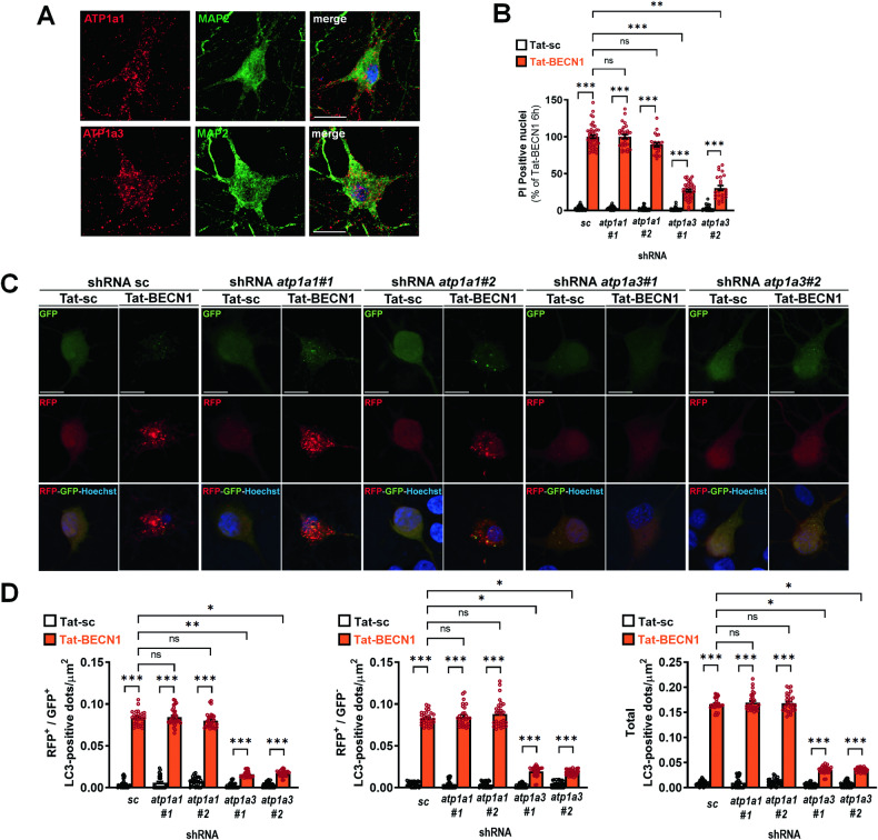
Macroautophagy (hereafter called autophagy) is an essential physiological process of degradation of organelles and long-lived proteins. The discovery of autosis, a Na/K-ATPase (ATP1)-dependent type of autophagic cell death with specific morphological and biochemical features, has strongly contributed to the acceptance of a pro-death role of autophagy. However, the occurrence and relevance of autosis in neurons has never been clearly investigated, whereas we previously provided evidence that autophagy mechanisms could be involved in neuronal death in different in vitro and in vivo rodent models of hypoxia-ischemia (HI) and that morphological features of autosis were observed in dying neurons following rat perinatal cerebral HI. In the present study, we demonstrated that neuronal autosis could occur in primary cortical neurons using two different stimulations enhancing autophagy flux and neuronal death: a neurotoxic concentration of Tat-BECN1 (an autophagy-inducing peptide) and a hypoxic/excitotoxic stimulus (mimicking neuronal death induced by cerebral HI). Both stimulations induce autophagic neuronal death (dependent on canonical autophagic genes and independent on apoptotic, necroptotic or ferroptotic pathways) with all morphological and biochemical (ATP1a-dependent) features of autosis. However, we demonstrated that autosis is not dependent on the ubiquitous subunit ATP1a1 in neurons, as in dividing cell types, but on the neuronal specific ATP1a3 subunit. We also provided evidence that, in different in vitro and in vivo models where autosis is induced, ATP1a3-BECN1 interaction is increased and prevented by cardiac glycosides treatment. Interestingly, an increase in ATP1a3-BECN1 interaction is also detected in dying neurons in the autoptic brains of human newborns with severe hypoxic-ischemic encephalopathy (HIE). Altogether, these results suggest that ATP1a3-BECN1-dependent autosis could play an important role in neuronal death in HI conditions, paving the way for the development of new neuroprotective strategies in hypoxic-ischemic conditions including in severe case of human HIE.
自噬(以下简称自噬)是降解细胞器和长寿命蛋白质的基本生理过程。自噬性细胞死亡的一种 Na/K-ATP 酶(ATP1)依赖性类型——自溶的发现,具有特定的形态和生化特征,强烈促进了自噬的促死亡作用的接受。然而,自溶在神经元中的发生和相关性从未被明确研究过,而我们之前提供的证据表明,自噬机制可能参与不同体外和体内缺氧-缺血(HI)的啮齿动物模型中的神经元死亡,并且在大鼠围产期脑 HI 后死亡的神经元中观察到自溶的形态特征。在本研究中,我们使用两种不同的刺激增强自噬通量和神经元死亡的方法,证明了原代皮质神经元中可能发生神经元自溶:一种神经毒性浓度的 Tat-BECN1(一种自噬诱导肽)和一种低氧/兴奋毒性刺激(模拟由脑 HI 引起的神经元死亡)。这两种刺激都诱导了具有自溶所有形态和生化(依赖于经典自噬基因且不依赖于凋亡、坏死或铁死亡途径)特征的自噬性神经元死亡。然而,我们证明了自溶不依赖于神经元中的普遍亚基 ATP1a1,而依赖于神经元特异性的 ATP1a3 亚基。我们还提供了证据表明,在不同的体外和体内模型中诱导自溶时,ATP1a3-BECN1 相互作用增加,并通过心脏糖苷治疗来预防。有趣的是,在 autoptic 脑中有严重缺氧缺血性脑病(HIE)的人类新生儿的死亡神经元中也检测到 ATP1a3-BECN1 相互作用增加。总之,这些结果表明,ATP1a3-BECN1 依赖性自溶可能在 HI 条件下的神经元死亡中发挥重要作用,为开发包括严重人类 HIE 在内的缺氧缺血条件下的新神经保护策略铺平了道路。